![Array Tomography Array Tomography]({"xsmall":"https://www.zeiss.com/content/dam/rms/reference-master/applications/life-sciences/volume-em/array-tomography/array-tomography.jpg/_jcr_content/renditions/original.image_file.100.56.file/array-tomography.jpg","small":"https://www.zeiss.com/content/dam/rms/reference-master/applications/life-sciences/volume-em/array-tomography/array-tomography.jpg/_jcr_content/renditions/original.image_file.360.203.file/array-tomography.jpg","medium":"https://www.zeiss.com/content/dam/rms/reference-master/applications/life-sciences/volume-em/array-tomography/array-tomography.jpg/_jcr_content/renditions/original.image_file.768.432.file/array-tomography.jpg","large":"https://www.zeiss.com/content/dam/rms/reference-master/applications/life-sciences/volume-em/array-tomography/array-tomography.jpg/_jcr_content/renditions/original.image_file.1024.576.file/array-tomography.jpg","xlarge":"https://www.zeiss.com/content/dam/rms/reference-master/applications/life-sciences/volume-em/array-tomography/array-tomography.jpg/_jcr_content/renditions/original.image_file.1280.720.file/array-tomography.jpg","xxlarge":"https://www.zeiss.com/content/dam/rms/reference-master/applications/life-sciences/volume-em/array-tomography/array-tomography.jpg/_jcr_content/renditions/original.image_file.1440.810.file/array-tomography.jpg","max":"https://www.zeiss.com/content/dam/rms/reference-master/applications/life-sciences/volume-em/array-tomography/array-tomography.jpg/_jcr_content/renditions/original./array-tomography.jpg"})
Volume EM
Array Tomography
標準仕様の走査電子顕微鏡を使用した非破壊ボリュームイメージング
ワークフローの略図
![試料調製 試料調製]({"xsmall":"https://www.zeiss.com/content/dam/rms/reference-master/applications/life-sciences/volume-em/array-tomography/array-tomography-sample-preparation.png/_jcr_content/renditions/original.image_file.100.100.file/array-tomography-sample-preparation.png","small":"https://www.zeiss.com/content/dam/rms/reference-master/applications/life-sciences/volume-em/array-tomography/array-tomography-sample-preparation.png/_jcr_content/renditions/original.image_file.360.360.file/array-tomography-sample-preparation.png","medium":"https://www.zeiss.com/content/dam/rms/reference-master/applications/life-sciences/volume-em/array-tomography/array-tomography-sample-preparation.png/_jcr_content/renditions/original.image_file.768.768.file/array-tomography-sample-preparation.png","large":"https://www.zeiss.com/content/dam/rms/reference-master/applications/life-sciences/volume-em/array-tomography/array-tomography-sample-preparation.png/_jcr_content/renditions/original.image_file.1024.1024.file/array-tomography-sample-preparation.png","xlarge":"https://www.zeiss.com/content/dam/rms/reference-master/applications/life-sciences/volume-em/array-tomography/array-tomography-sample-preparation.png/_jcr_content/renditions/original./array-tomography-sample-preparation.png","xxlarge":"https://www.zeiss.com/content/dam/rms/reference-master/applications/life-sciences/volume-em/array-tomography/array-tomography-sample-preparation.png/_jcr_content/renditions/original./array-tomography-sample-preparation.png","max":"https://www.zeiss.com/content/dam/rms/reference-master/applications/life-sciences/volume-em/array-tomography/array-tomography-sample-preparation.png/_jcr_content/renditions/original./array-tomography-sample-preparation.png"})
1
樹脂包埋試料を切削し、切片を並べます。各切片の厚さは通常30~70 nmで、切削順に切片を試料キャリアに貼り付けます。
![画像取得 画像取得]({"xsmall":"https://www.zeiss.com/content/dam/rms/reference-master/applications/life-sciences/volume-em/array-tomography/array-tomography-image-acquisition.png/_jcr_content/renditions/original.image_file.100.100.file/array-tomography-image-acquisition.png","small":"https://www.zeiss.com/content/dam/rms/reference-master/applications/life-sciences/volume-em/array-tomography/array-tomography-image-acquisition.png/_jcr_content/renditions/original.image_file.360.360.file/array-tomography-image-acquisition.png","medium":"https://www.zeiss.com/content/dam/rms/reference-master/applications/life-sciences/volume-em/array-tomography/array-tomography-image-acquisition.png/_jcr_content/renditions/original.image_file.768.768.file/array-tomography-image-acquisition.png","large":"https://www.zeiss.com/content/dam/rms/reference-master/applications/life-sciences/volume-em/array-tomography/array-tomography-image-acquisition.png/_jcr_content/renditions/original.image_file.1024.1024.file/array-tomography-image-acquisition.png","xlarge":"https://www.zeiss.com/content/dam/rms/reference-master/applications/life-sciences/volume-em/array-tomography/array-tomography-image-acquisition.png/_jcr_content/renditions/original./array-tomography-image-acquisition.png","xxlarge":"https://www.zeiss.com/content/dam/rms/reference-master/applications/life-sciences/volume-em/array-tomography/array-tomography-image-acquisition.png/_jcr_content/renditions/original./array-tomography-image-acquisition.png","max":"https://www.zeiss.com/content/dam/rms/reference-master/applications/life-sciences/volume-em/array-tomography/array-tomography-image-acquisition.png/_jcr_content/renditions/original./array-tomography-image-acquisition.png"})
2
連続する各切片を、走査電子顕微鏡(SEM)でイメージングします。
![セグメンテーションの処理 セグメンテーションの処理]({"xsmall":"https://www.zeiss.com/content/dam/rms/reference-master/applications/life-sciences/volume-em/array-tomography/volume-em-processing-segmentation.png/_jcr_content/renditions/original.image_file.100.100.file/volume-em-processing-segmentation.png","small":"https://www.zeiss.com/content/dam/rms/reference-master/applications/life-sciences/volume-em/array-tomography/volume-em-processing-segmentation.png/_jcr_content/renditions/original.image_file.360.360.file/volume-em-processing-segmentation.png","medium":"https://www.zeiss.com/content/dam/rms/reference-master/applications/life-sciences/volume-em/array-tomography/volume-em-processing-segmentation.png/_jcr_content/renditions/original.image_file.768.768.file/volume-em-processing-segmentation.png","large":"https://www.zeiss.com/content/dam/rms/reference-master/applications/life-sciences/volume-em/array-tomography/volume-em-processing-segmentation.png/_jcr_content/renditions/original.image_file.1024.1024.file/volume-em-processing-segmentation.png","xlarge":"https://www.zeiss.com/content/dam/rms/reference-master/applications/life-sciences/volume-em/array-tomography/volume-em-processing-segmentation.png/_jcr_content/renditions/original./volume-em-processing-segmentation.png","xxlarge":"https://www.zeiss.com/content/dam/rms/reference-master/applications/life-sciences/volume-em/array-tomography/volume-em-processing-segmentation.png/_jcr_content/renditions/original./volume-em-processing-segmentation.png","max":"https://www.zeiss.com/content/dam/rms/reference-master/applications/life-sciences/volume-em/array-tomography/volume-em-processing-segmentation.png/_jcr_content/renditions/original./volume-em-processing-segmentation.png"})
3
取得した電子顕微鏡画像が処理され、デジタルに調整されて3Dデータセットになります。細胞コンパートメントを特定し、セグメント化することができます。
![3Dビジュアライゼーション解析 3Dビジュアライゼーション解析]({"xsmall":"https://www.zeiss.com/content/dam/rms/reference-master/applications/life-sciences/volume-em/array-tomography/volume-em-3d-visualization-analysis.png/_jcr_content/renditions/original.image_file.100.100.file/volume-em-3d-visualization-analysis.png","small":"https://www.zeiss.com/content/dam/rms/reference-master/applications/life-sciences/volume-em/array-tomography/volume-em-3d-visualization-analysis.png/_jcr_content/renditions/original.image_file.360.360.file/volume-em-3d-visualization-analysis.png","medium":"https://www.zeiss.com/content/dam/rms/reference-master/applications/life-sciences/volume-em/array-tomography/volume-em-3d-visualization-analysis.png/_jcr_content/renditions/original.image_file.768.768.file/volume-em-3d-visualization-analysis.png","large":"https://www.zeiss.com/content/dam/rms/reference-master/applications/life-sciences/volume-em/array-tomography/volume-em-3d-visualization-analysis.png/_jcr_content/renditions/original.image_file.1024.1024.file/volume-em-3d-visualization-analysis.png","xlarge":"https://www.zeiss.com/content/dam/rms/reference-master/applications/life-sciences/volume-em/array-tomography/volume-em-3d-visualization-analysis.png/_jcr_content/renditions/original./volume-em-3d-visualization-analysis.png","xxlarge":"https://www.zeiss.com/content/dam/rms/reference-master/applications/life-sciences/volume-em/array-tomography/volume-em-3d-visualization-analysis.png/_jcr_content/renditions/original./volume-em-3d-visualization-analysis.png","max":"https://www.zeiss.com/content/dam/rms/reference-master/applications/life-sciences/volume-em/array-tomography/volume-em-3d-visualization-analysis.png/_jcr_content/renditions/original./volume-em-3d-visualization-analysis.png"})
4
セグメント化された3Dデータセットは、視覚化および調査を行い、統計的に分析することができます。
アプリケーション例
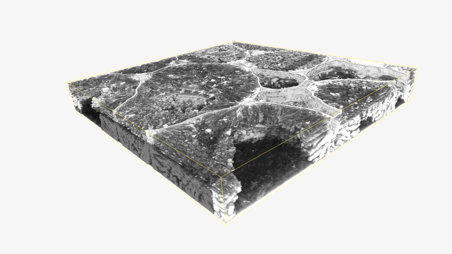
プラスモデスマータが分布した根粒から作製された連続切片の3D再構成
![植物と細菌の共生関係 植物と細菌の共生関係]({"xsmall":"https://www.zeiss.com/content/dam/rms/reference-master/applications/life-sciences/volume-em/array-tomography/root-nodules-correlative-array-tomography.jpg/_jcr_content/renditions/original.image_file.100.100.file/root-nodules-correlative-array-tomography.jpg","small":"https://www.zeiss.com/content/dam/rms/reference-master/applications/life-sciences/volume-em/array-tomography/root-nodules-correlative-array-tomography.jpg/_jcr_content/renditions/original.image_file.360.360.file/root-nodules-correlative-array-tomography.jpg","medium":"https://www.zeiss.com/content/dam/rms/reference-master/applications/life-sciences/volume-em/array-tomography/root-nodules-correlative-array-tomography.jpg/_jcr_content/renditions/original.image_file.768.768.file/root-nodules-correlative-array-tomography.jpg","large":"https://www.zeiss.com/content/dam/rms/reference-master/applications/life-sciences/volume-em/array-tomography/root-nodules-correlative-array-tomography.jpg/_jcr_content/renditions/original./root-nodules-correlative-array-tomography.jpg","xlarge":"https://www.zeiss.com/content/dam/rms/reference-master/applications/life-sciences/volume-em/array-tomography/root-nodules-correlative-array-tomography.jpg/_jcr_content/renditions/original./root-nodules-correlative-array-tomography.jpg","xxlarge":"https://www.zeiss.com/content/dam/rms/reference-master/applications/life-sciences/volume-em/array-tomography/root-nodules-correlative-array-tomography.jpg/_jcr_content/renditions/original./root-nodules-correlative-array-tomography.jpg","max":"https://www.zeiss.com/content/dam/rms/reference-master/applications/life-sciences/volume-em/array-tomography/root-nodules-correlative-array-tomography.jpg/_jcr_content/renditions/original./root-nodules-correlative-array-tomography.jpg"})
![植物と細菌の共生関係 ご提供:D. Sherrier, J. Caplan, and S. Modla, University of Delaware, USA 植物と細菌の共生関係 ご提供:D. Sherrier, J. Caplan, and S. Modla, University of Delaware, USA]({"xsmall":"https://www.zeiss.com/content/dam/rms/reference-master/applications/life-sciences/volume-em/array-tomography/root-nodules-correlative-array-tomography.jpg/_jcr_content/renditions/original.image_file.100.100.file/root-nodules-correlative-array-tomography.jpg","small":"https://www.zeiss.com/content/dam/rms/reference-master/applications/life-sciences/volume-em/array-tomography/root-nodules-correlative-array-tomography.jpg/_jcr_content/renditions/original.image_file.360.360.file/root-nodules-correlative-array-tomography.jpg","medium":"https://www.zeiss.com/content/dam/rms/reference-master/applications/life-sciences/volume-em/array-tomography/root-nodules-correlative-array-tomography.jpg/_jcr_content/renditions/original.image_file.768.768.file/root-nodules-correlative-array-tomography.jpg","large":"https://www.zeiss.com/content/dam/rms/reference-master/applications/life-sciences/volume-em/array-tomography/root-nodules-correlative-array-tomography.jpg/_jcr_content/renditions/original./root-nodules-correlative-array-tomography.jpg","xlarge":"https://www.zeiss.com/content/dam/rms/reference-master/applications/life-sciences/volume-em/array-tomography/root-nodules-correlative-array-tomography.jpg/_jcr_content/renditions/original./root-nodules-correlative-array-tomography.jpg","xxlarge":"https://www.zeiss.com/content/dam/rms/reference-master/applications/life-sciences/volume-em/array-tomography/root-nodules-correlative-array-tomography.jpg/_jcr_content/renditions/original./root-nodules-correlative-array-tomography.jpg","max":"https://www.zeiss.com/content/dam/rms/reference-master/applications/life-sciences/volume-em/array-tomography/root-nodules-correlative-array-tomography.jpg/_jcr_content/renditions/original./root-nodules-correlative-array-tomography.jpg"})
ご提供:D. Sherrier, J. Caplan, and S. Modla, University of Delaware, USA
植物と細菌の共生関係
植物の健康状態に対する根粒内の細菌の影響を理解する
植物の根は、ネットワークを張り巡らせることで、あらゆる植物の成長に欠かせない水や栄養のすべての源にアクセスします。植物の健康や産出量を最適化するには、根のネットワーク全体を調べ、外部の微生物の影響を理解することが重要です。植物と根粒内の細菌における共生関係を調べるにあたっては、根粒と細菌分布に関する知識が必要であり、これを詳細に理解するためには、蛍光および高分解能の構造評価を組み合わせることが必須となります。
Correlative Array Tomographyでは蛍光および構造データにおける両方のオーバーレイが可能なため、根粒と細菌分布を視覚化することができます。