

Array Tomographyのアプリケーション
革新的な事例を見るArray Tomography(AT)は、ナノスケールの分解能で3Dデータセットを生成する強力なアプローチです。試料は数百の切片にセクショニングされ、各切片が走査電子顕微鏡を使用して高分解能でイメージングされます。
非破壊的な方法として、試料の超薄切片をイメージング用基板上に置き、蛍光顕微鏡法などの手法やその他の解析アプローチを用いて観察します。
ボリュームEM(vEM)法には特別な装置が必要ないため、走査電子顕微鏡(SEM)とウルトラミクロトームがあればどのようなラボでもボリュームEMを活用できます。このような特徴を備えたATは、例からも分かるように、細胞から組織、植物に至るまで多様な試料の観察に有益なアプローチです。
脳組織の神経ネットワークを理解する
数百万の神経ネットワークを調べることで、脳のシグナル伝達経路に関する理解が深まります。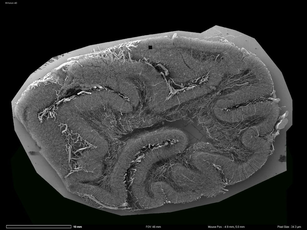

ATを用いて取得したサルの脳のオーバービュー画像。
ATを用いて取得したサルの脳のオーバービュー画像。
脳は、数百万の神経ネットワークとシグナル伝達経路を有する複雑な器官です。神経組織の構造と機能との関係を理解することは、この複雑さを部分的に解明するのに役立ち、脳の働きを深く理解することにつながるだけでなく、長期的な機能不全の医療介入による治療法の解明にも役立ちます。

SEMを用いて取得したサルの脳の高解像度画像。
SEMを用いて取得したサルの脳の高解像度画像。
数百万の神経ネットワークを調べるには、高分解能3Dイメージングが必要です。小さな脳の試料では、全体の3Dデータセットを取得するのに時間がかかりますが可能な範囲です。ただし、マウスやサルの脳などの非常に大きな組織の場合は、イメージングをスケールアップさせて、現実的な時間枠の中で高分解能3Dデータセットを生成する必要があります。ATなら、これを実現できます。


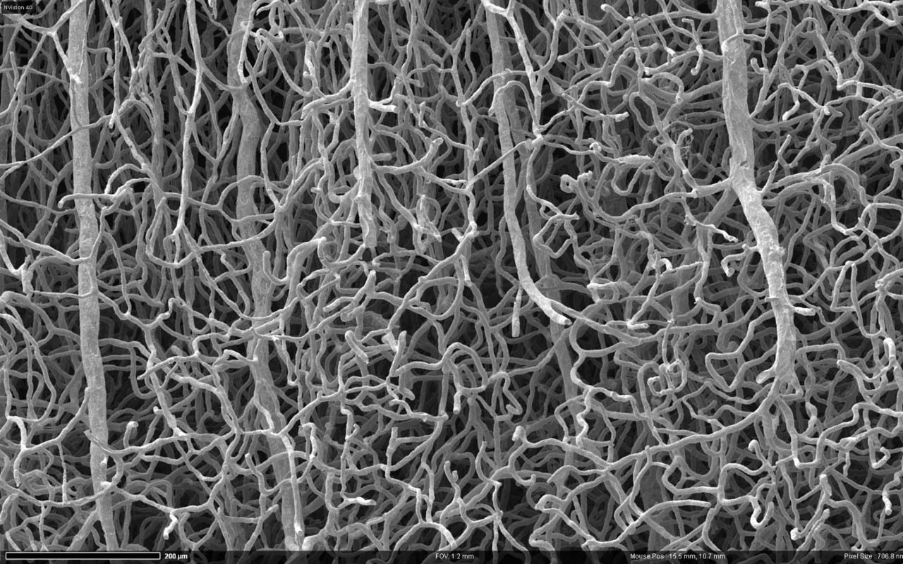
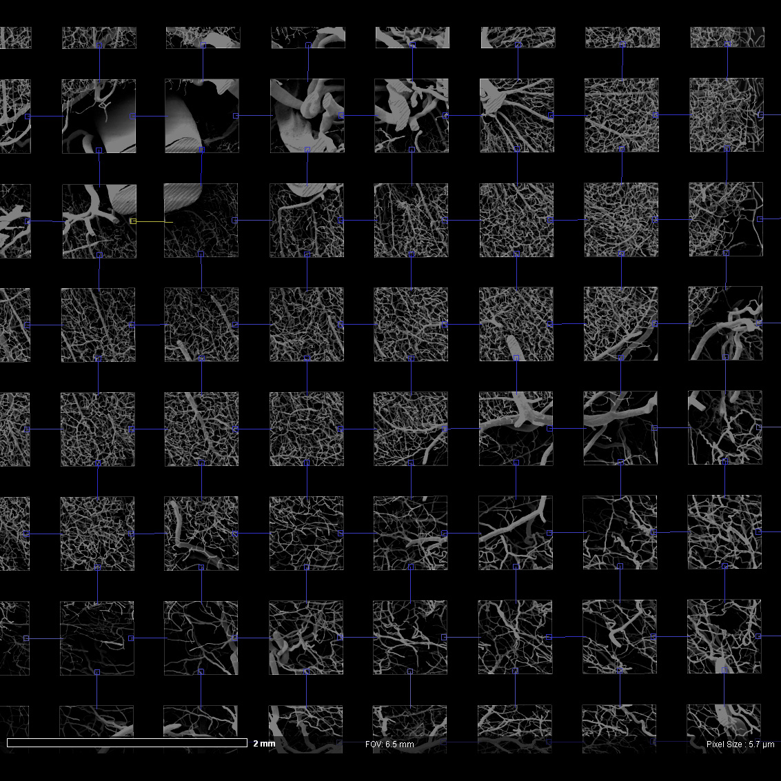
ATを用いて取得したサルの脳血管。
ATを用いて取得したサルの脳血管。
ZEISS Atlas 5 Array Tomographyは、基板上の連続する切片をナノメーターレベルの分解能で自動的にイメージングするハードウェア/ソフトウェアツールです。この独自のワークフローは使いやすいだけでなく、特に自動イメージング用に設計されており、脳組織の膨大な結合を解明するのに必要となる大容量の3Dビジュアライゼーションを可能にします。

自動で取得した、脳血管を示すサルの脳の拡大画像。Atlas 5 Array Tomographyを用いて取得した画像。実視野:3700 mm
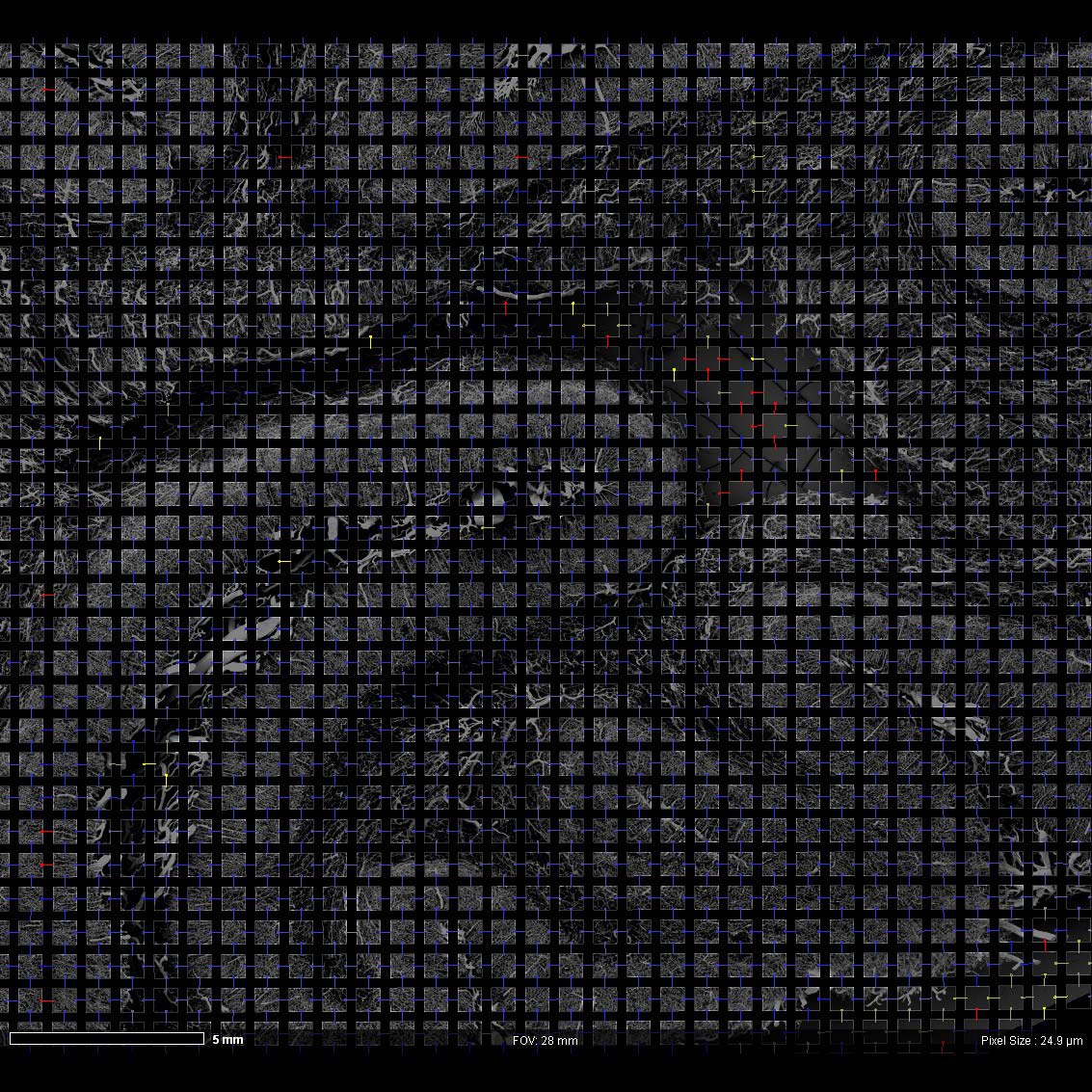

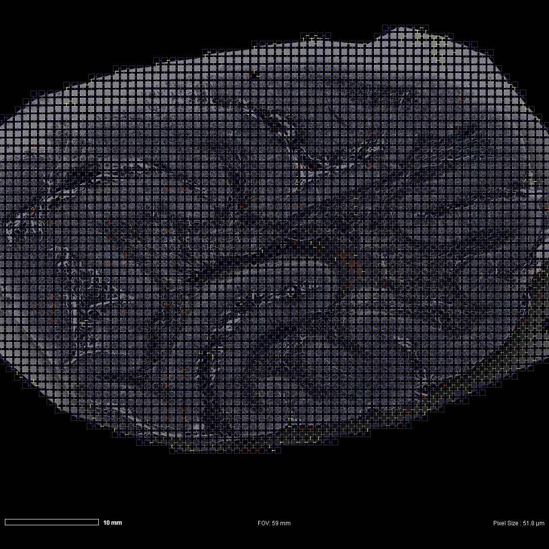
1000を超える画像をつなぎ合わせたサルの脳を表すモザイク画像。各タイル画像は4096 x 4096ピクセル(ピクセルサイズ150 nm)。

タイル画像の自動スティッチングにより作成した、サルの脳の広視野画像。


マウス脳の超薄切片


マウス脳の超薄切片。試料ご提供:J. Lichtman, Harvard University, USA
マウス脳の超薄切片。試料ご提供:J. Lichtman, Harvard University, USA
Atlas 5 Array Tomographyのコンピューター支援ツールを使うことにより、何百もの連続する切片上におけるあらゆる形状の関心領域を制限なく定義できます。これは特に、サルまたはマウスの脳の神経ネットワークを捉える実験等で役立ちます。


マウス脳の超薄切片
マウス脳の超薄切片。試料ご提供:J. Lichtman, Harvard University, USA
マウス脳の超薄切片。試料ご提供:J. Lichtman, Harvard University, USA
ここに示すのは、基板に載せたマウス脳の超薄切片の拡大SEM画像です。画像の自動取得をサポートするAtlas 5 Array Tomographyによって撮影されました。
核やミトコンドリアなどの細胞内の詳細構造が特定でき、さらに3Dで再構成されると、異なるニューロン間の結合がマッピングされます。


マウス脳の超薄切片


マウス脳の超薄切片。試料ご提供:J. Lichtman, Harvard University, USA
マウス脳の超薄切片。試料ご提供:J. Lichtman, Harvard University, USA
この画像は、テープでマウントされ、イメージングの準備ができた多数の脳切片を示しています。Atlas 5 Array Tomographyは、後の高分解能SEMによる取得のために小脳の各切片を特定し、各切片の有益な情報すべてをソフトウェアプラットフォームに保存します。
この動画は、Atlas 5 Array Tomographyを使用してATのワークフローを効率化することがもたらす大きな価値を示しています。動画が進むにつれて、各切片から取得された高分解能データがどのように組み合わされ、最終的に3Dデータセットを生成するかを見ることができます。


マウス脳の超薄切片
高分解能SEMで視神経を捉えるには、各切片をイメージングしてその後再構成する必要があります。Array Tomographyは各切片の位置を自動で特定し、その後イメージングするため、迅速でシンプルな取得プロセスが実現します。


マウス脳の超薄切片
この画像は、Atlas 5ソフトウェアを使用してイメージングする前の、ウェハ上に載せられた視神経の各切片を示しています。
Array Tomographyを用いて脳をイメージングする際は、数百、あるいは数千もの切片の位置を特定してイメージングする必要があります。この切片をそれぞれ手動で特定するのは大変な作業であり、あまりにも時間がかかります。Atlas 5は、各切片を自動で素早く特定するため、その後のイメージングを素早く効率的に開始でき、各切片のすべての高分解能情報がソフトウェアに登録されます。
植物の健康状態に対する根粒内の細菌の影響を理解する
Correlative Array Tomography(CAT)を用いた蛍光および構造データのオーバーレイにより、根粒と細菌の分布を視覚化できます。

プラスモデスマータの分布を示す根粒


プラスモデスマータの分布を示す根粒。ご提供:D. Sherrier, J. Caplan, and S. Modla, University of Delaware, USA
プラスモデスマータの分布を示す根粒。ご提供:D. Sherrier, J. Caplan, and S. Modla, University of Delaware, USA
植物の根は、ネットワークを張り巡らせることで、すべての植物の成長に欠かせない水や栄養にアクセスします。植物の健康や産出量を最適化するには、根のネットワーク全体を調べ、外部の微生物の影響を理解することが重要です。
植物と根粒内の細菌における共生関係を調べるにあたっては、根粒と細菌分布に関する知識が必要であり、これを詳細に理解するためには、蛍光および高分解能の構造評価を組み合わせることが必須となります。
Correlative Array Tomographyでは蛍光および構造データにおける両方のオーバーレイが可能なため、根粒と細菌分布を視覚化することができます。
根粒内の細菌が植物の状態に与える影響をきちんと理解するためには、高分解能の構造情報を精度の高い蛍光情報と組み合わせることが重要です。
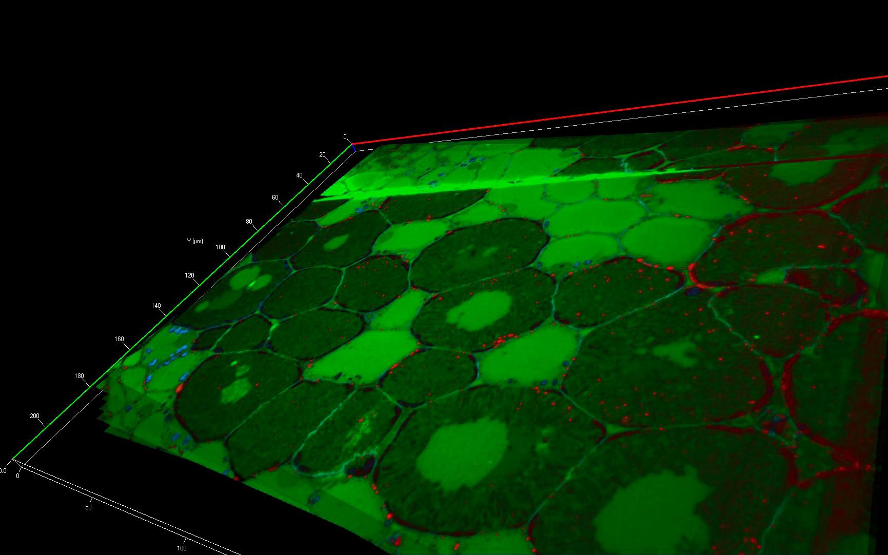

プラスモデスマータの分布を示す根粒。ご提供:D. Sherrier, J. Caplan, and S. Modla, University of Delaware, USA
これらの画像は、根粒の連続する切片から得られた蛍光データの3D再構成または1枚の切片であり、プラスモデスマータの分布を示しています。SEMデータを用いてスタックをオーバーレイすることで、細菌感染とプラスモデスマータの分布との関係を調べることができます。
試料はEponで包埋し、ウルトラミクロトームで切断しています。連続する切片を、マイクロマニピュレーターでITOコーティングしたカバーガラスに移動しました。細胞壁はCalcofluor White(青)で、プラスモデスマータはカロースに対してAlexa Fluor 647で染色されました。SEMでのイメージングのため、試料は後染色されています。


プラスモデスマータの分布を示す根粒


プラスモデスマータの分布を示す根粒


プラスモデスマータの分布を示す根粒


プラスモデスマータの分布を示す根粒


プラスモデスマータの分布を示す根粒


プラスモデスマータの分布を示す根粒


Array Tomographyによる画像取得の設定方法
この動画では、ZEISS Atlas 5 Array Tomographyを使用して、蛍光およびSEMデータとの相関データセットを生成するワークフローを説明します。動画内で示される例の1つは酵母試料で、この酵母の構成を示しています。
動画内の酵母試料はEponで包埋後、ウルトラミクロトームで切断されています。連続する切片を、マイクロマニピュレーターでITOコーティングしたカバーガラスに移動しました。試料はまず光学顕微鏡でイメージングされ、その後SEMによるイメージングのために後染色されました。
ご提供:D. Sherrier, J. Caplan, and S. Modla, University of Delaware, USA
ハンチントン病の疾患生物学と進行を理解する
Correlative Array Tomography(CAT)を用いてマクロファージ内のタンパク質プラークの発達を視覚化できます。

変異型ハンチンチンタンパク質の凝集により誘発されたタンパク質プラークを示すマクロファージ
変異型ハンチンチンタンパク質の凝集により誘発されたタンパク質プラークを示すマクロファージ。この画像は、1つの切片からの蛍光データセットで、マクロファージ内のタンパク質プラークを示しています。DNA:青(DAPI)、ハンチンチンタンパク質:赤(Alexa Fluor 647)。ご提供:Jeff Caplan, University of Delaware, USA
変異型ハンチンチンタンパク質の凝集により誘発されたタンパク質プラークを示すマクロファージ。この画像は、1つの切片からの蛍光データセットで、マクロファージ内のタンパク質プラークを示しています。DNA:青(DAPI)、ハンチンチンタンパク質:赤(Alexa Fluor 647)。ご提供:Jeff Caplan, University of Delaware, USA
ハンチントン病は、ハンチンチンタンパク質をコーディングする4番染色体上の遺伝子の欠陥によって生じる不治で進行性の神経変性疾患です。欠陥遺伝子は変異型ハンチンチンタンパク質の形成をもたらします。変異型ハンチンチンタンパク質はミスフォールドして凝集し、脳内でタンパク質プラークの発達を引き起こします。このような脳の変質により、運動、行動、思考、感情の混乱が生じます。


ハンチンチンの過剰発現の結果、凝集したタンパク質がZスタック内で明確に視認できる


ハンチンチンの過剰発現の結果、凝集したタンパク質がZスタック内で明確に視認できます。GFP-Huntington抗体を使用して、細胞内のハンチンチンタンパク質のプラークの位置を特定しました(Alexa Fluor 647、赤)。核は青で示されています(Hoechst)。ハンチンチンプラークの分布と核の位置を3Dで示すLMおよびSEMのZスタックの相関。ご提供:J. Caplan, E. Kmiec and S. Modla, University of Delaware, USA
ハンチンチンの過剰発現の結果、凝集したタンパク質がZスタック内で明確に視認できます。GFP-Huntington抗体を使用して、細胞内のハンチンチンタンパク質のプラークの位置を特定しました(Alexa Fluor 647、赤)。核は青で示されています(Hoechst)。ハンチンチンプラークの分布と核の位置を3Dで示すLMおよびSEMのZスタックの相関。ご提供:J. Caplan, E. Kmiec and S. Modla, University of Delaware, USA
マクロファージ内でのタンパク質プラークの発達(ハンチントン病の研究のモデル系)を視覚化することと、これらが微細構造の細胞細部にどのように関連しているかを理解することは、ハンチントン病の疾患生物学と進行の理解において重要です。
Correlative Array Tomographyにより、このマクロファージの連続切片から微細構造と蛍光データの両方を取得することができます。


ハンチンチンプラークの分布と核の位置を3Dで示すLMおよびSEMのZスタックの相関
ハンチンチンプラークの分布と核の位置を3Dで示すLMおよびSEMのZスタックの相関。ご提供:J. Caplan, E. Kmiec and S. Modla, University of Delaware, USA
ハンチンチンプラークの分布と核の位置を3Dで示すLMおよびSEMのZスタックの相関。ご提供:J. Caplan, E. Kmiec and S. Modla, University of Delaware, USA
ハンチントン病の疾患生物学と進行をきちんと理解するには、微細構造および蛍光データの相関が重要です。
左の画像は、ハンチンチンタンパク質を発現しているマクロファージの連続LM画像とSEM画像を組み合わせて3Dで取得した情報を示しています。画像は、ZEISS ZEN Correlative Array Tomographyを用いた実験で取得しました。


