![シリアルブロックフェイス走査電子顕微鏡法を用いたVolume EM シリアルブロックフェイス走査電子顕微鏡法を用いたVolume EM]({"xsmall":"https://www.zeiss.com/content/dam/rms/reference-master/applications/life-sciences/volume-em/serial-block-face-sem/serial-block-face-sem.jpg/_jcr_content/renditions/original.image_file.100.56.file/serial-block-face-sem.jpg","small":"https://www.zeiss.com/content/dam/rms/reference-master/applications/life-sciences/volume-em/serial-block-face-sem/serial-block-face-sem.jpg/_jcr_content/renditions/original.image_file.360.203.file/serial-block-face-sem.jpg","medium":"https://www.zeiss.com/content/dam/rms/reference-master/applications/life-sciences/volume-em/serial-block-face-sem/serial-block-face-sem.jpg/_jcr_content/renditions/original.image_file.768.432.file/serial-block-face-sem.jpg","large":"https://www.zeiss.com/content/dam/rms/reference-master/applications/life-sciences/volume-em/serial-block-face-sem/serial-block-face-sem.jpg/_jcr_content/renditions/original.image_file.1024.576.file/serial-block-face-sem.jpg","xlarge":"https://www.zeiss.com/content/dam/rms/reference-master/applications/life-sciences/volume-em/serial-block-face-sem/serial-block-face-sem.jpg/_jcr_content/renditions/original.image_file.1280.720.file/serial-block-face-sem.jpg","xxlarge":"https://www.zeiss.com/content/dam/rms/reference-master/applications/life-sciences/volume-em/serial-block-face-sem/serial-block-face-sem.jpg/_jcr_content/renditions/original.image_file.1440.810.file/serial-block-face-sem.jpg","max":"https://www.zeiss.com/content/dam/rms/reference-master/applications/life-sciences/volume-em/serial-block-face-sem/serial-block-face-sem.jpg/_jcr_content/renditions/original./serial-block-face-sem.jpg"})
シリアルブロックフェイスSEM
高度に自動化されたセクショニングとボリュームデータイメージング
ワークフローの略図
![シリアルブロックフェイスSEMイメージング シリアルブロックフェイスSEMイメージング]({"xsmall":"https://www.zeiss.com/content/dam/rms/reference-master/applications/life-sciences/volume-em/serial-block-face-sem/serial-block-face-sem-imaging.png/_jcr_content/renditions/original.image_file.100.100.file/serial-block-face-sem-imaging.png","small":"https://www.zeiss.com/content/dam/rms/reference-master/applications/life-sciences/volume-em/serial-block-face-sem/serial-block-face-sem-imaging.png/_jcr_content/renditions/original.image_file.360.360.file/serial-block-face-sem-imaging.png","medium":"https://www.zeiss.com/content/dam/rms/reference-master/applications/life-sciences/volume-em/serial-block-face-sem/serial-block-face-sem-imaging.png/_jcr_content/renditions/original.image_file.768.768.file/serial-block-face-sem-imaging.png","large":"https://www.zeiss.com/content/dam/rms/reference-master/applications/life-sciences/volume-em/serial-block-face-sem/serial-block-face-sem-imaging.png/_jcr_content/renditions/original.image_file.1024.1024.file/serial-block-face-sem-imaging.png","xlarge":"https://www.zeiss.com/content/dam/rms/reference-master/applications/life-sciences/volume-em/serial-block-face-sem/serial-block-face-sem-imaging.png/_jcr_content/renditions/original./serial-block-face-sem-imaging.png","xxlarge":"https://www.zeiss.com/content/dam/rms/reference-master/applications/life-sciences/volume-em/serial-block-face-sem/serial-block-face-sem-imaging.png/_jcr_content/renditions/original./serial-block-face-sem-imaging.png","max":"https://www.zeiss.com/content/dam/rms/reference-master/applications/life-sciences/volume-em/serial-block-face-sem/serial-block-face-sem-imaging.png/_jcr_content/renditions/original./serial-block-face-sem-imaging.png"})
1
SEMチャンバー内に配置されたウルトラミクロトームを使用して、樹脂包埋試料を切断します。露出した試料表面をイメージングします。この切断とイメージングのプロセスを、目的の構造が完全にイメージングできるまで繰り返し行います。
![セグメンテーションの処理 セグメンテーションの処理]({"xsmall":"https://www.zeiss.com/content/dam/rms/reference-master/applications/life-sciences/volume-em/array-tomography/volume-em-processing-segmentation.png/_jcr_content/renditions/original.image_file.100.100.file/volume-em-processing-segmentation.png","small":"https://www.zeiss.com/content/dam/rms/reference-master/applications/life-sciences/volume-em/array-tomography/volume-em-processing-segmentation.png/_jcr_content/renditions/original.image_file.360.360.file/volume-em-processing-segmentation.png","medium":"https://www.zeiss.com/content/dam/rms/reference-master/applications/life-sciences/volume-em/array-tomography/volume-em-processing-segmentation.png/_jcr_content/renditions/original.image_file.768.768.file/volume-em-processing-segmentation.png","large":"https://www.zeiss.com/content/dam/rms/reference-master/applications/life-sciences/volume-em/array-tomography/volume-em-processing-segmentation.png/_jcr_content/renditions/original.image_file.1024.1024.file/volume-em-processing-segmentation.png","xlarge":"https://www.zeiss.com/content/dam/rms/reference-master/applications/life-sciences/volume-em/array-tomography/volume-em-processing-segmentation.png/_jcr_content/renditions/original./volume-em-processing-segmentation.png","xxlarge":"https://www.zeiss.com/content/dam/rms/reference-master/applications/life-sciences/volume-em/array-tomography/volume-em-processing-segmentation.png/_jcr_content/renditions/original./volume-em-processing-segmentation.png","max":"https://www.zeiss.com/content/dam/rms/reference-master/applications/life-sciences/volume-em/array-tomography/volume-em-processing-segmentation.png/_jcr_content/renditions/original./volume-em-processing-segmentation.png"})
2
取得した電子顕微鏡画像が処理され、デジタルに調整されて3Dデータセットになります。細胞コンパートメントを特定し、セグメント化することができます。
![3Dビジュアライゼーション解析 3Dビジュアライゼーション解析]({"xsmall":"https://www.zeiss.com/content/dam/rms/reference-master/applications/life-sciences/volume-em/array-tomography/volume-em-3d-visualization-analysis.png/_jcr_content/renditions/original.image_file.100.100.file/volume-em-3d-visualization-analysis.png","small":"https://www.zeiss.com/content/dam/rms/reference-master/applications/life-sciences/volume-em/array-tomography/volume-em-3d-visualization-analysis.png/_jcr_content/renditions/original.image_file.360.360.file/volume-em-3d-visualization-analysis.png","medium":"https://www.zeiss.com/content/dam/rms/reference-master/applications/life-sciences/volume-em/array-tomography/volume-em-3d-visualization-analysis.png/_jcr_content/renditions/original.image_file.768.768.file/volume-em-3d-visualization-analysis.png","large":"https://www.zeiss.com/content/dam/rms/reference-master/applications/life-sciences/volume-em/array-tomography/volume-em-3d-visualization-analysis.png/_jcr_content/renditions/original.image_file.1024.1024.file/volume-em-3d-visualization-analysis.png","xlarge":"https://www.zeiss.com/content/dam/rms/reference-master/applications/life-sciences/volume-em/array-tomography/volume-em-3d-visualization-analysis.png/_jcr_content/renditions/original./volume-em-3d-visualization-analysis.png","xxlarge":"https://www.zeiss.com/content/dam/rms/reference-master/applications/life-sciences/volume-em/array-tomography/volume-em-3d-visualization-analysis.png/_jcr_content/renditions/original./volume-em-3d-visualization-analysis.png","max":"https://www.zeiss.com/content/dam/rms/reference-master/applications/life-sciences/volume-em/array-tomography/volume-em-3d-visualization-analysis.png/_jcr_content/renditions/original./volume-em-3d-visualization-analysis.png"})
3
セグメント化された3Dデータセットは、視覚化および調査を行い、統計的に分析することができます。
アプリケーション例
構造と機能との関係を理解する
![ウルトラミクロトーム内蔵のZEISS Sigmaを使用して、7 nmピクセル、75枚の画像スタックでイメージングされたマウスの脳。ミクロトームは切片ごとに15 nm切り取るよう設定。 ウルトラミクロトーム内蔵のZEISS Sigmaを使用して、7 nmピクセル、75枚の画像スタックでイメージングされたマウスの脳。ミクロトームは切片ごとに15 nm切り取るよう設定。]({"xsmall":"https://www.zeiss.com/content/dam/rms/reference-master/applications/life-sciences/volume-em/serial-block-face-sem/mouse-brain-sbf-sem-sigma-3view.jpg/_jcr_content/renditions/original.image_file.100.100.file/mouse-brain-sbf-sem-sigma-3view.jpg","small":"https://www.zeiss.com/content/dam/rms/reference-master/applications/life-sciences/volume-em/serial-block-face-sem/mouse-brain-sbf-sem-sigma-3view.jpg/_jcr_content/renditions/original.image_file.360.360.file/mouse-brain-sbf-sem-sigma-3view.jpg","medium":"https://www.zeiss.com/content/dam/rms/reference-master/applications/life-sciences/volume-em/serial-block-face-sem/mouse-brain-sbf-sem-sigma-3view.jpg/_jcr_content/renditions/original.image_file.768.768.file/mouse-brain-sbf-sem-sigma-3view.jpg","large":"https://www.zeiss.com/content/dam/rms/reference-master/applications/life-sciences/volume-em/serial-block-face-sem/mouse-brain-sbf-sem-sigma-3view.jpg/_jcr_content/renditions/original./mouse-brain-sbf-sem-sigma-3view.jpg","xlarge":"https://www.zeiss.com/content/dam/rms/reference-master/applications/life-sciences/volume-em/serial-block-face-sem/mouse-brain-sbf-sem-sigma-3view.jpg/_jcr_content/renditions/original./mouse-brain-sbf-sem-sigma-3view.jpg","xxlarge":"https://www.zeiss.com/content/dam/rms/reference-master/applications/life-sciences/volume-em/serial-block-face-sem/mouse-brain-sbf-sem-sigma-3view.jpg/_jcr_content/renditions/original./mouse-brain-sbf-sem-sigma-3view.jpg","max":"https://www.zeiss.com/content/dam/rms/reference-master/applications/life-sciences/volume-em/serial-block-face-sem/mouse-brain-sbf-sem-sigma-3view.jpg/_jcr_content/renditions/original./mouse-brain-sbf-sem-sigma-3view.jpg"})
ウルトラミクロトーム内蔵のZEISS Sigmaを使用して、7 nmピクセル、75枚の画像スタックでイメージングされたマウスの脳。ミクロトームは切片ごとに15 nm切り取るよう設定。
ニューロンにおける微細構造の詳細なイメージング
脳は、数百万の神経ネットワークとシグナル伝達経路を有する複雑な器官です。脳組織の構造と機能との関係を理解することは、この複雑さを部分的に解明するのに役立ち、神経ネットワークの構造と機能を深く理解することにつながり、長期的には特定の疾患の医療介入による治療法の解明にも役立ちます。
![Focal Charge Compensation装置を使用して、2.5 keV、画素滞在時間1 μsピクセル、高真空でイメージングしたブロックフェース試料。スケールバー:1 µm。ご提供:NCMIR Focal Charge Compensation装置を使用して、2.5 keV、画素滞在時間1 μsピクセル、高真空でイメージングしたブロックフェース試料。スケールバー:1 µm。ご提供:NCMIR]({"xsmall":"https://www.zeiss.com/content/dam/rms/reference-master/applications/life-sciences/volume-em/serial-block-face-sem/cultured-hippocampal-neurons.jpg/_jcr_content/renditions/original.image_file.100.100.file/cultured-hippocampal-neurons.jpg","small":"https://www.zeiss.com/content/dam/rms/reference-master/applications/life-sciences/volume-em/serial-block-face-sem/cultured-hippocampal-neurons.jpg/_jcr_content/renditions/original.image_file.360.360.file/cultured-hippocampal-neurons.jpg","medium":"https://www.zeiss.com/content/dam/rms/reference-master/applications/life-sciences/volume-em/serial-block-face-sem/cultured-hippocampal-neurons.jpg/_jcr_content/renditions/original.image_file.768.768.file/cultured-hippocampal-neurons.jpg","large":"https://www.zeiss.com/content/dam/rms/reference-master/applications/life-sciences/volume-em/serial-block-face-sem/cultured-hippocampal-neurons.jpg/_jcr_content/renditions/original./cultured-hippocampal-neurons.jpg","xlarge":"https://www.zeiss.com/content/dam/rms/reference-master/applications/life-sciences/volume-em/serial-block-face-sem/cultured-hippocampal-neurons.jpg/_jcr_content/renditions/original./cultured-hippocampal-neurons.jpg","xxlarge":"https://www.zeiss.com/content/dam/rms/reference-master/applications/life-sciences/volume-em/serial-block-face-sem/cultured-hippocampal-neurons.jpg/_jcr_content/renditions/original./cultured-hippocampal-neurons.jpg","max":"https://www.zeiss.com/content/dam/rms/reference-master/applications/life-sciences/volume-em/serial-block-face-sem/cultured-hippocampal-neurons.jpg/_jcr_content/renditions/original./cultured-hippocampal-neurons.jpg"})
Focal Charge Compensation装置を使用して、2.5 keV、画素滞在時間1 μsピクセル、高真空でイメージングしたブロックフェース試料。スケールバー:1 µm。ご提供:NCMIR
細胞培養中のニューロンのイメージング
SBF-SEMは、樹状突起や軸索など、神経細胞における細長く突出した構造を連続的にイメージングするのに最適なソリューションです。細胞培養中のニューロンのイメージングは特に困難です。なぜなら、非導電樹脂の割合が高く、試料が帯電しやすくなるからです。Focal Charge Compensationは帯電効果を低減し、高画質でのイメージングを実現します。SBF-SEMとFocal Charge Compensationとを組み合わせることで、ニューロンの微細構造の細部も容易に分解およびイメージングできます。
これらの画像は、シナプス後膜肥厚(矢印)を染色したPSD95-APEX2を示す、培養された海馬ニューロンの3Dデータの1枚の切片です。画像はZEISS FESEM、内蔵されたウルトラミクロトーム、Focal Charge Compensationを使用し取得しました。帯電効果の除去によって達成された高分解能イメージングにより、薄い樹状突起や結合などの微細構造が視認できます。
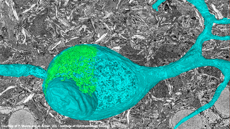
マウス脳組織内の単一のニューロンと細胞コンパートメント
この動画は、シリアルブロックフェイスSEMを用いて取得したマウス脳試料の断面を示しています。それぞれのシングルブロックフェース画像から、このアプローチで得られる分解能の高さがはっきりとわかります。Z方向に並んでいる単一のニューロン、および細胞コンパートメントが特定できます。