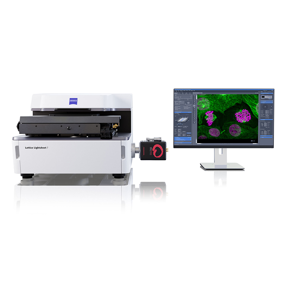
ZEISS Lattice Lightsheet 7
Product

ZEISS Lattice Lightsheet 7

Long-term volumetric imaging of living cells

ZEISS Lattice Lightsheet 7 makes light sheet fluorescence microscopy available for live cell imaging at subcellular resolution – while also allowing you to use your standard sample carriers. With this automated, easy-to-use system, volumetric imaging of subcellular structures and dynamics over hours and days with best protection from photo damage becomes available to everyone. Discover the dynamics of life in unprecedented depth of detail – with the ease you never imagined possible!

  • Examine living specimens directly on your standard sample carriers
  • Watch the subcellular dynamics of life over hours and even days
  • Reveal three-dimensional details in their true proportions
  • Don’t miss an interesting event on your coverslip
  • Use single molecule microscopy to investigate structures or dynamics at the nanometer scale

Discover the subcellular dynamics of life
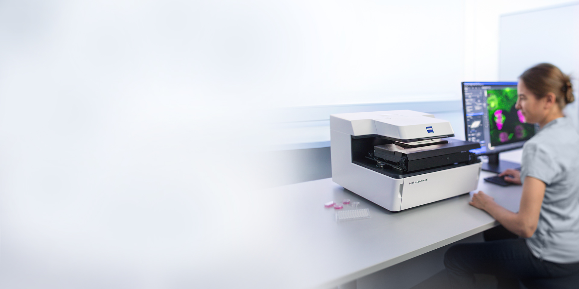
  • ZEISS Lattice Lightsheet 7 - Workflow

Lattice Light Sheet Technology Made Accessible to Everyone

Discover the dynamics of life in unprecedented depth of detail – with the ease you never imagined possible!

Light sheet fluorescence microscopy has become the method of choice for fast and gentle imaging of living specimens. Lattice Lightsheet 7 enables live cell imaging at subcellular resolution, by using standard sample carriers you already use for confocal microscopy.

With this automated, easy-to-use system the benefit of lattice light sheet microscopy – volumetric imaging of subcellular structures and dynamics over hours and days with best protection from photo damage – becomes available to everyone.

Virtually no phototoxicity or photobleaching

Observe the dynamics of life at subcellular resolution and follow how the finest structures evolve over time. Conventional imaging systems often become too invasive, causing photodamage that limits long-term live-cell experiments. ZEISS Lattice Lightsheet 7 overcomes these limitations with lattice-structured illumination that automatically adapts to the sensitivity of your sample, dramatically reducing photobleaching and phototoxicity. This allows you to image living specimens continuously for hours or even days. A precisely controlled incubation environment, combined with an integrated auto-immersion mechanism, supports reliable, unattended long-term imaging.

U2OS cell expressing Lifeact-tdTomato, color-coded depth projection, recorded for 5.5 days over 4 mitotic cycles

High-speed volumetric imaging

The extremely fast image acquisition of ZEISS Lattice Lightsheet 7 enables up to three volume scans per second. Dynamic imaging of full sample volumes with this high temporal resolution means no longer missing an interesting event on your coverslip. Near-isotropic resolution along the X, Y and Z axes gives you a three-dimensional image of your sample that reveals structural details in their true proportions. Fast laser switching allows for imaging using up to three colors practically simultaneously, with minimized color crosstalk.

Video: COS-7 cell transiently transfected with Tomm20-mEmerald and Calreticulin-tdTomato. The example shows ER wrapping around mitochondria and assisting mitochondrial fission.

From the earliest stages to self-organization

Lattice light-sheet microscopy provides a gentle window into life’s earliest processes, enabling researchers to observe development from the first cell divisions to complex tissue organization without disrupting delicate biological dynamics.

Mouse preimplantation embryo (1-2 cell stage) expressing H2B-mCherry and 2xmNeonGreen-CENP-C to visualize chromosomes and kinetochores, respectively.
Image courtesy: Manami Koshiguchi, RIKEN Center for Biosystems Dynamics Research, Kobe, Japan

  • LLC-PK1 cell undergoing mitosis. Cells are expressing H2B-mCherry (orange) and a-Tubulin mEGFP (cyan-to-yellow).
  • Unique insights into the relationship between dynamics and structure

    ZEISS Lattice Lightsheet 7 combines gentle live-cell imaging with Single Molecule Localization Microscopy (SMLM) to reveal how cellular dynamics and molecular structures are connected. Cells can be monitored in real time, fixed at a defined state, and subsequently analyzed at molecular resolution. With ultra-thin lattice light sheets delivering high signal-to-noise ratios and precise optical sectioning, the system enables visualization of even single molecules across entire cells.

    Due to the angled view and inherent optical sectioning, Lattice Lightsheet 7 allows for imaging whole cells with molecular detail. Depending on theorientation, subcellular compartments can be visualized with down to 10 nm localization precision.

    LLC-PK1 cell undergoing mitosis. Cells are expressing H2B-mCherry (orange) and a-Tubulin mEGFP (cyan-to-yellow).

  • Unique insights into the relationship between dynamics and structure

    ZEISS Lattice Lightsheet 7 combines gentle live-cell imaging with Single Molecule Localization Microscopy (SMLM) to reveal how cellular dynamics and molecular structures are connected. Cells can be monitored in real time, fixed at a defined state, and subsequently analyzed at molecular resolution. With ultra-thin lattice light sheets delivering high signal-to-noise ratios and precise optical sectioning, the system enables visualization of even single molecules across entire cells.

    Due to the angled view and inherent optical sectioning, Lattice Lightsheet 7 allows for imaging whole cells with molecular detail. Depending on theorientation, subcellular compartments can be visualized with down to 10 nm localization precision.

    LLC-PK1 cell undergoing mitosis. Cells are expressing H2B-mCherry (orange) and a-Tubulin mEGFP (cyan-to-yellow).

  • Unique insights into the relationship between dynamics and structure

    ZEISS Lattice Lightsheet 7 combines gentle live-cell imaging with Single Molecule Localization Microscopy (SMLM) to reveal how cellular dynamics and molecular structures are connected. Cells can be monitored in real time, fixed at a defined state, and subsequently analyzed at molecular resolution. With ultra-thin lattice light sheets delivering high signal-to-noise ratios and precise optical sectioning, the system enables visualization of even single molecules across entire cells.

    Due to the angled view and inherent optical sectioning, Lattice Lightsheet 7 allows for imaging whole cells with molecular detail. Depending on theorientation, subcellular compartments can be visualized with down to 10 nm localization precision.

    LLC-PK1 cell undergoing mitosis. Cells are expressing H2B-mCherry (orange) and a-Tubulin mEGFP (cyan-to-yellow).

  • Unique insights into the relationship between dynamics and structure

    ZEISS Lattice Lightsheet 7 combines gentle live-cell imaging with Single Molecule Localization Microscopy (SMLM) to reveal how cellular dynamics and molecular structures are connected. Cells can be monitored in real time, fixed at a defined state, and subsequently analyzed at molecular resolution. With ultra-thin lattice light sheets delivering high signal-to-noise ratios and precise optical sectioning, the system enables visualization of even single molecules across entire cells.

    Due to the angled view and inherent optical sectioning, Lattice Lightsheet 7 allows for imaging whole cells with molecular detail. Depending on theorientation, subcellular compartments can be visualized with down to 10 nm localization precision.

    LLC-PK1 cell undergoing mitosis. Cells are expressing H2B-mCherry (orange) and a-Tubulin mEGFP (cyan-to-yellow).

  • Spheroids formed from U2OS cells expressing Actin-EGFP, infiltrating the extracellular matrix (collagen), imaged for 24hrs every 5min. Color-coded depth projection
  • 3D cell culture models

    Lattice Light Sheet Microscopy provides gentle, high-speed 3D imaging using structured light sheets to minimize phototoxicity and photobleaching. 3D cell culture models, including spheroids and organoids, better recapitulate tissue architecture and gradients than 2D cultures, enabling more physiologically relevant studies.

  • Spheroids formed from U2OS cells expressing Actin-EGFP, infiltrating the extracellular matrix (collagen), imaged for 24hrs every 5min. Color-coded depth projection
  • Whole tissue sections

    Lattice Light Sheet Microscopy enables rapid, high-resolution 3D imaging with minimal phototoxicity by using a structured light sheet. For fast imaging of whole tissue sections, this approach can scan large volumes quickly while preserving cellular detail and tissue context.

    ZEISS Lattice Lightsheet 7 combines fast volumetric imaging with high resolution to capture large and complex samples, including tissue sections and expanded specimens. Its ability to image entire volumes efficiently while preserving image quality enables comprehensive insights into how biological structures and complexity emerge over time.

  • Spheroids formed from U2OS cells expressing Actin-EGFP, infiltrating the extracellular matrix (collagen), imaged for 24hrs every 5min. Color-coded depth projection
  • Single Molecule Localization Microscopy (SMLM)

    Single Molecule Localization Microscopy (SMLM) creates ultra-high-resolution images by detecting individual fluorescent molecules one at a time. By precisely locating each molecule and repeating the process millions of times, it builds a highly detailed image at the nanoscale. One widely used SMLM method is STORM, which uses stochastic activation of fluorophores to localize single molecules with nanometer precision. Each binding event produces a detectable signal that reveals the target's exact location. Together, these repeated events generate exceptionally detailed images of cellular structures.

  • Spheroids formed from U2OS cells expressing Actin-EGFP, infiltrating the extracellular matrix (collagen), imaged for 24hrs every 5min. Color-coded depth projection
  • Structures and dynamics as complex as life itself

    For oncology and drug discovery researchers, ZEISS Lattice Lightsheet 7 provides robust, physiologically relevant insights into invasion mechanisms under near-native conditions. By capturing key behaviors linked to metastatic potential—such as the active invasion of tumor spheroids into their extracellular matrix over extended periods—the system enables a deeper understanding of cancer progression. Its gentle lattice illumination preserves cell viability while continuously imaging complex 3D cell culture models, revealing dynamic processes such as coordinated cell migration and cell–cell organization. With ZEISS Lattice Lightsheet 7, researchers can explore the intricate structures and behaviors that define living systems with unprecedented temporal and spatial detail.

    Spheroids formed from U2OS cells expressing Actin-EGFP, infiltrating the extracellular matrix (collagen), imaged for 24hrs every 5min. Color-coded depth projection

  • Structures and dynamics as complex as life itself

    For oncology and drug discovery researchers, ZEISS Lattice Lightsheet 7 provides robust, physiologically relevant insights into invasion mechanisms under near-native conditions. By capturing key behaviors linked to metastatic potential—such as the active invasion of tumor spheroids into their extracellular matrix over extended periods—the system enables a deeper understanding of cancer progression. Its gentle lattice illumination preserves cell viability while continuously imaging complex 3D cell culture models, revealing dynamic processes such as coordinated cell migration and cell–cell organization. With ZEISS Lattice Lightsheet 7, researchers can explore the intricate structures and behaviors that define living systems with unprecedented temporal and spatial detail.

    Spheroids formed from U2OS cells expressing Actin-EGFP, infiltrating the extracellular matrix (collagen), imaged for 24hrs every 5min. Color-coded depth projection

Product features

Incubation chamber loaded with a standard 35 mm dish.

Incubation chamber loaded with a standard 35 mm dish.

Standard sample carriers usage

Without having to adapt your usual sample preparation, you can examine living specimens directly on the sample carriers you already use for confocal microscopy. ZEISS Lattice Lightsheet 7 can be used with all standard sample carriers that come with a no. 1.5 coverslip for the bottom:
  • Slides
  • 35 mm dishes
  • Chamber slides
  • Multi-well plates
ZEISS Lattice Lightsheet 7 - LED Illumination

Gentle transmission illumination ensures that your sample is quickly located.

Fast and gentle sample location

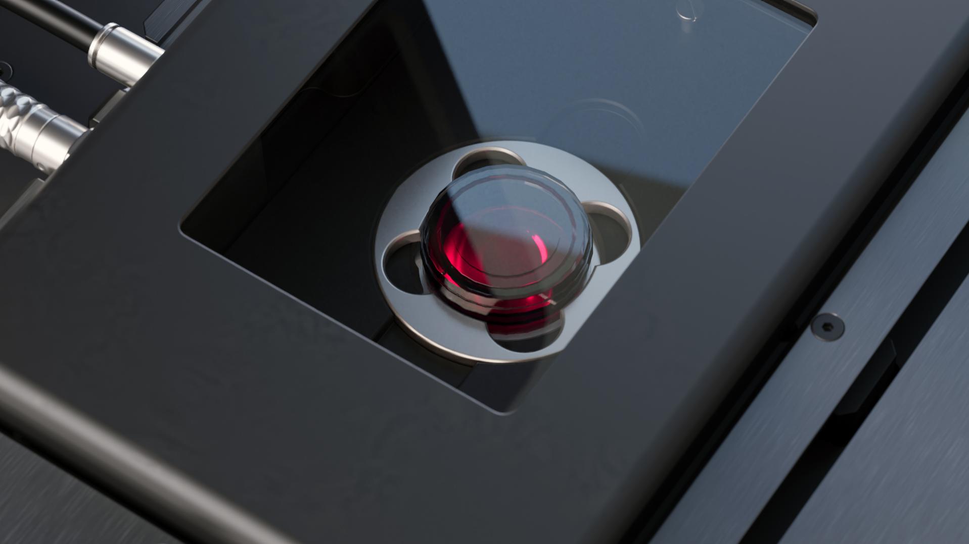
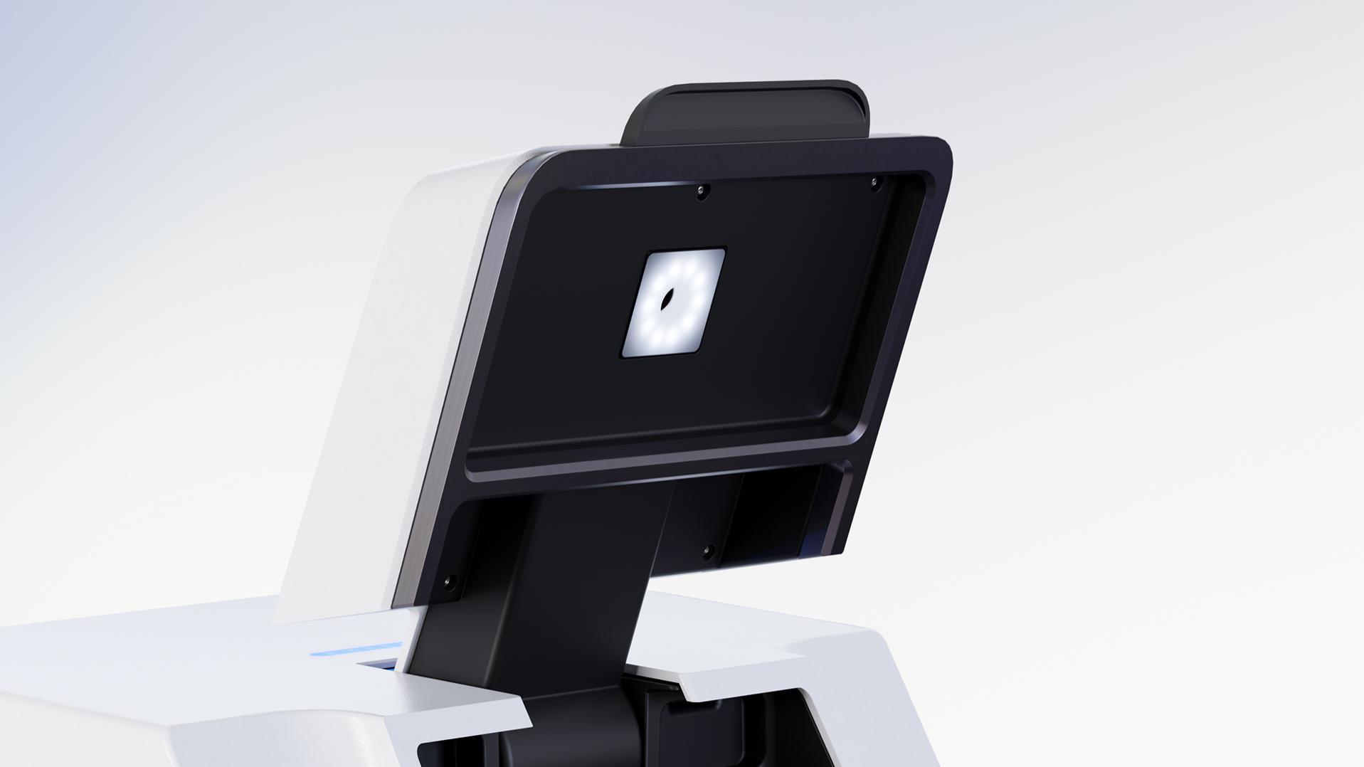
With the integrated transmission LEDs and oblique detection which provide a DIC-like contrast, you can easily locate your sample. Change from white to red transmission LEDs for more gentle illumination if necessary. And you can choose to include transmitted light illumination during long-term observations.
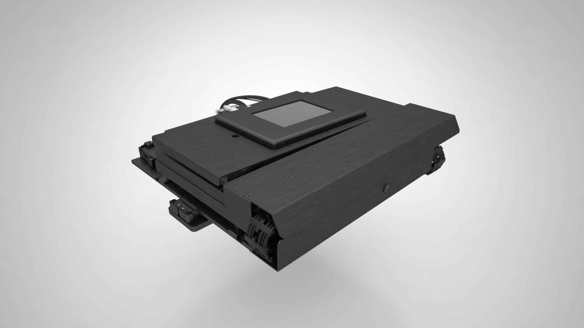
ZEISS Lattice Lightsheet 7 - 5-axis Stage

The 5-axis stage combines highest precision and speed with a large travel range for multiwell plate imaging.

Automatic sample leveling

Specifically designed for this system, the unique 5-axis stage not only allows movement along the X, Y and Z axes, but also tilting with the highest precision in X and Y, compensating for even the smallest deviations in carrier dimensions or sample position. Leveling your sample is done automatically, which relieves you of tedious manual procedures.
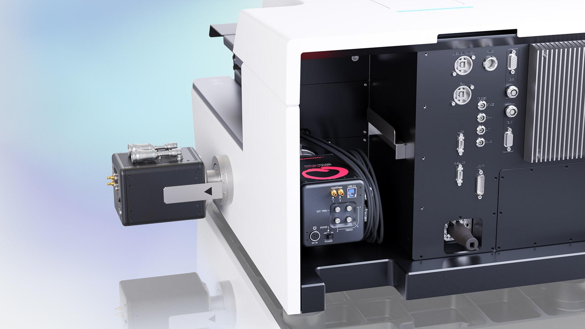
ZEISS Lattice Lightsheet 7 - Dual-Camera Configuration

Dual-camera configuration of ZEISS Lattice Lightsheet 7.

Doubled temporal resolution with two cameras

The innovative design of the excitation beam path allows simultaneous excitation of the sample with multiple laser lines. Combined with two Hamamatsu ORCA-Fusion cameras, this enables truly simultaneous imaging of two channels, which is critical for a range of applications such as ratiometric experiments. A dual-camera setup also allows you to use single bandpass filters in front of each camera to minimize crosstalk and achieve cleanest results without compromising speed.
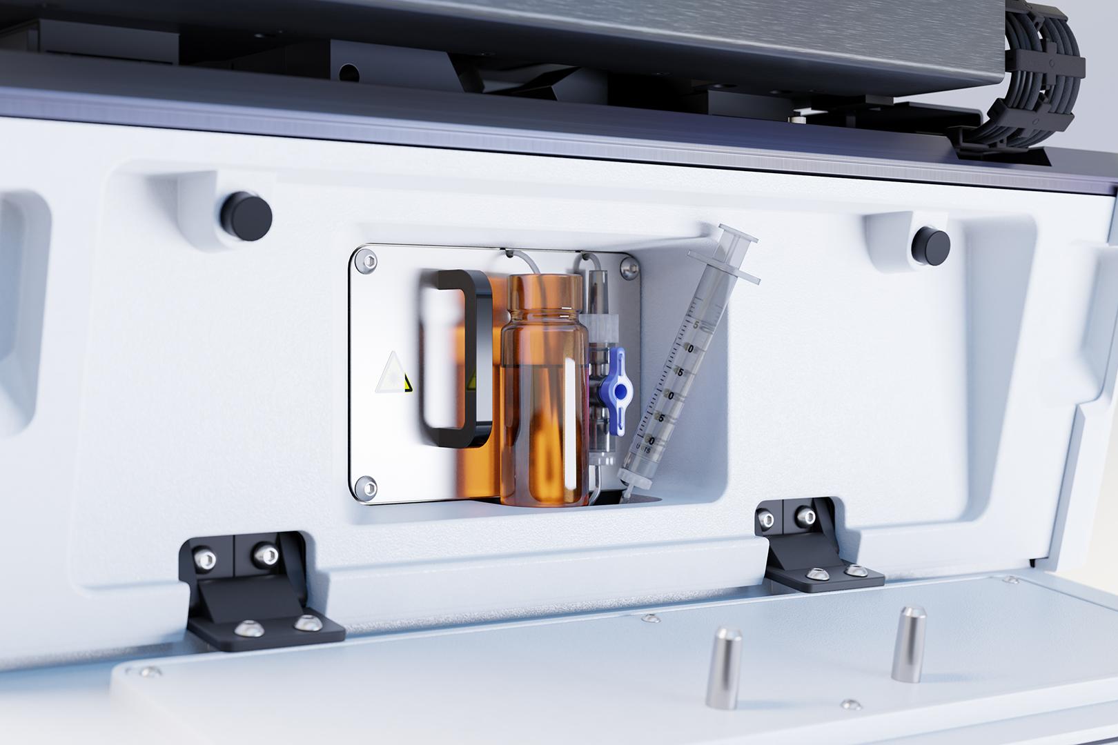
ZEISS Lattice Lightsheet 7 - Autoimmersion

ZEISS Lattice Lightsheet 7 autoimmersion equipment.

Unattended long-term experiments

Incubation: An integrated incubation system provides long-term stability throughout varying environmental conditions. The microscope controls and monitors temperature, CO2 and O2 levels, and humidity automatically, to preserve the integrity of your sample throughout the experiments. The lid with glass window allows quick and easy access to the sample to facilitate its inspection between experimental runs.

Autoimmersion: Prime the system to release any air, then a supply of immersion media tailored to the needs of your experiments is released automatically. Replenishing the immersion media is software-controlled, so you don’t have to worry about interfering with image acquisition. The reservoir is protected from illumination to keep bacterial growth at bay. Objectives are shielded from immersion supply; hence they remain dry, even if excess immersion media is applied.
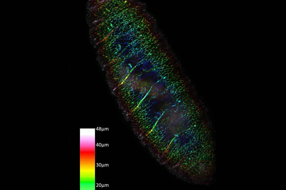
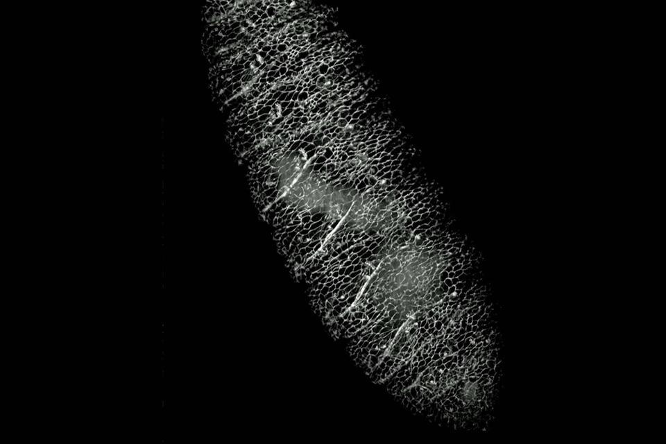
  • Color-coded depth projection of a Drosphila embryo with GPF labeling.
  • This video shows a drosophila embryo with GFP labeling as it moves over time. A total of 91,100 i mages were taken, 911 volume planes, 100 time points. One volume, every 15 secs; imaging duration 25 mins, imaging volume: 300 × 455 × 145 μm3.

Drosophila embryo

Drosophila melanogaster is a model organism in many research fields such as biomedical research. Many genetically modified variants are available to researchers. This video shows a drosophila embryo with GFP labeling as it moves over time. A total of 91,100 images were taken, 911 volume planes, 100 time points. One volume, every 15 secs; imaging duration 25 mins, imaging volume: 300 × 455 × 145 μm3.

Drug discovery and development

  • Cell proliferation

    Cell proliferation is a fundamental process for understanding tissue growth and disease progression, particularly in cancer. Studying its underlying mechanisms enables researchers to develop targeted therapies that help control abnormal cell growth.

    Image courtesy: Abel Pereira da Graca, ZMCC Oberkochen, Germany

  • T-cell mechanism & efficacy

    T cells play a central role in the immune response by recognizing and eliminating infected or cancerous cells. Research into their mechanisms and function is driving the development of more effective immunotherapies, helping improve treatment outcomes for a wide range of diseases.

    Image courtesy: Elisa Sanchez, Morgan Huse lab, Memorial Sloan Kettering Cancer Center, New York

  • Scratch assay

    Wound healing assays provide a model for studying tissue repair and evaluating treatment effectiveness. By replicating the wound closure process, they enable researchers to investigate healing mechanisms and advance the development of therapies for skin injuries and related conditions.

    Image courtesy: Abel Pereira da Graca, ZMCC Oberkochen, Germany

  • Antibody uptake assays

    Studying antibody uptake in 4D provides dynamic insights into antibody–cell interactions in a physiologically relevant 3D environment. Unlike endpoint assays, it distinguishes true cellular uptake from membrane retention and enables precise quantification over time. Lattice Lightsheet 7 helps optimize therapeutic antibody uptake, supporting improved drug design, delivery, and treatment efficacy.

    4D time series of a U2OS cell from the start of exposure to Trastuzumab until 48hrs later, when antibody uptake had reached saturation. Trastuzumab was labelled with pHrodo Red. Arivis Pro was used to segment compartments with Trastuzumab uptake and color-coded for fluorescence intensity.

  • Antibody uptake assays

    Studying antibody uptake in 4D provides dynamic insights into antibody–cell interactions in a physiologically relevant 3D environment. Unlike endpoint assays, it distinguishes true cellular uptake from membrane retention and enables precise quantification over time. Lattice Lightsheet 7 helps optimize therapeutic antibody uptake, supporting improved drug design, delivery, and treatment efficacy.

    3D rendering of U2OS cell after 24hrs of exposure to Trastuzumab labelled with pHrodo Red (cyan) and Zenon Alexa647 (orange). pHrodo Red fluoresces in acidic environments such as endosomes and lysosomes, which are the intracellular compartments where internalized antibodies are trafficked.

  • Antibody uptake assays

    Studying antibody uptake in 4D provides dynamic insights into antibody–cell interactions in a physiologically relevant 3D environment. Unlike endpoint assays, it distinguishes true cellular uptake from membrane retention and enables precise quantification over time. Lattice Lightsheet 7 helps optimize therapeutic antibody uptake, supporting improved drug design, delivery, and treatment efficacy.

    Trastuzumab was labelled with pHrodo and added to U2OS cells. Antibody uptake was observed for 24hrs every 10min. Fluorescence signal was segmented in arivis Pro and color-coded for fluorescence intensity. IgG antibodies labelled with pHrodo served as negative control. Graphic illustrating Trastuzumab uptake over the period of 48hrs. Analysed with arivis Pro.

  • T-cell efficacy assays

    Lattice Lightsheet 7 enables real-time visualization of T cell dynamics, interactions, and immune responses, providing deeper insights into T cell function and therapeutic potential. Combined with multi-well plate compatibility, it supports screening and identification of promising genetically retargeted T cell candidates for next-generation immunotherapies.

    T cells are stained with a calcium dye. Cancer cells are stained with Lifeact-mCherry, which appear red under the imaging light. Cells shown in pink indicate apoptosis.

    Image courtesy of Ms. Kylie Luong, A/Prof Misty Jenkins, Niall Geoghegan, WEHI, Melbourne.

  • Antibody uptake

    The uptake of antibodies by cancer cells plays an important role in the effectiveness of antibody-based therapies for cancer and infectious diseases. Understanding these mechanisms can help optimize drug delivery and improve therapeutic outcomes in clinical applications.

  • T-cell efficacy assays

    Lattice Lightsheet 7 enables real-time visualization of T cell dynamics, interactions, and immune responses, providing deeper insights into T cell function and therapeutic potential. Combined with multi-well plate compatibility, it supports screening and identification of promising genetically retargeted T cell candidates for next-generation immunotherapies.

    Cytotoxic T-Lymphocyte interacting with a mouse melanoma target cell. T-Lymphocyte is stained with Cell Vue Maroon (orange) and the target cell is expessing F-tractin EGFP (green)

    Image courtesy: Elisa Sanchez, Morgan Huse lab, Memorial Sloan Kettering Cancer Center, New York

  • T-cell efficacy assays

    Lattice Lightsheet 7 enables real-time visualization of T cell dynamics, interactions, and immune responses, providing deeper insights into T cell function and therapeutic potential. Combined with multi-well plate compatibility, it supports screening and identification of promising genetically retargeted T cell candidates for next-generation immunotherapies.

    Cytotoxic T-Lymphocyte interacting with a mouse melanoma target cell. T-Lymphocyte is stained with Cell Vue Maroon (orange) and the target cell is expessing F-tractin EGFP (green)

    Image courtesy: Elisa Sanchez, Morgan Huse lab, Memorial Sloan Kettering Cancer Center, New York

  • T-cell efficacy assays

    Lattice Lightsheet 7 enables real-time visualization of T cell dynamics, interactions, and immune responses, providing deeper insights into T cell function and therapeutic potential. Combined with multi-well plate compatibility, it supports screening and identification of promising genetically retargeted T cell candidates for next-generation immunotherapies.

    Cytotoxic T-Lymphocyte interacting with a mouse melanoma target cell. T-Lymphocyte is stained with Cell Vue Maroon (orange) and the target cell is expessing F-tractin EGFP (green)

    Image courtesy: Elisa Sanchez, Morgan Huse lab, Memorial Sloan Kettering Cancer Center, New York

  • Cell proliferation assays

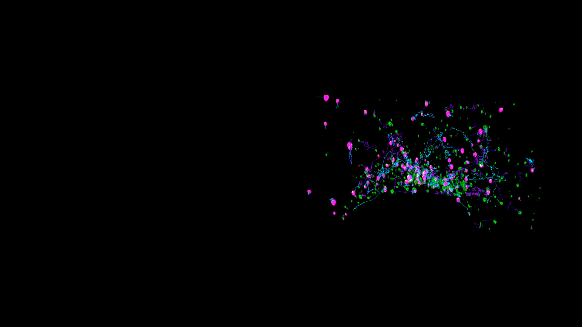
    Lattice Lightsheet 7 enables detailed 3D visualization of cell proliferation over time, revealing how cells interact, organize, migrate, and differentiate in response to stimuli. Combined with Arivis Pro’s AI-powered segmentation and tracking, researchers can automatically identify cells and nuclei, detect mitotic events, and follow cell divisions to distinguish mother and daughter cells, providing deeper insights into disease mechanisms and therapeutic development.

    LLC-PK1 cells undergoing mitosis. Cells are expressing H2B-mCherry (magenta) and α-Tubulin mEGFP (white).

    Data Acquisition and Analysis: Abel Pereira da Graca and Noemi Castroviejo Jimenez, ZM Customer Center Oberkochen, Germany.

  • Cell proliferation assays

    Lattice Lightsheet 7 enables detailed 3D visualization of cell proliferation over time, revealing how cells interact, organize, migrate, and differentiate in response to stimuli. Combined with Arivis Pro’s AI-powered segmentation and tracking, researchers can automatically identify cells and nuclei, detect mitotic events, and follow cell divisions to distinguish mother and daughter cells, providing deeper insights into disease mechanisms and therapeutic development.

    LLC-PK1 cells undergoing mitosis. Cells are expressing H2B-mCherry (magenta) and α-Tubulin mEGFP (white).

    Data Acquisition and Analysis: Abel Pereira da Graca and Noemi Castroviejo Jimenez, ZM Customer Center Oberkochen, Germany.

  • Cell proliferation assays

    Lattice Lightsheet 7 enables detailed 3D visualization of cell proliferation over time, revealing how cells interact, organize, migrate, and differentiate in response to stimuli. Combined with Arivis Pro’s AI-powered segmentation and tracking, researchers can automatically identify cells and nuclei, detect mitotic events, and follow cell divisions to distinguish mother and daughter cells, providing deeper insights into disease mechanisms and therapeutic development.

    LLC-PK1 cells undergoing mitosis. Cells are expressing H2B-mCherry (magenta) and α-Tubulin mEGFP (white).

    Data Acquisition and Analysis: Abel Pereira da Graca and Noemi Castroviejo Jimenez, ZM Customer Center Oberkochen, Germany.

  • Wound healing assays

    Lattice Lightsheet 7 enables real-time 3D visualization of cell migration and wound healing, capturing large volumes of dynamic cellular activity with high precision. This approach provides deeper insights into the mechanisms driving tissue repair by revealing how cells interact with their environment and respond during healing processes.

    LLC-PK1 cells undergoing mitosis. Cells are expressing H2B-mCherry (magenta) and α-Tubulin mEGFP (white).

    Data Acquisition and Analysis: Abel Pereira da Graca and Noemi Castroviejo Jimenez, ZM Customer Center Oberkochen, Germany.

  • Wound healing assays

    Lattice Lightsheet 7 enables real-time 3D visualization of cell migration and wound healing, capturing large volumes of dynamic cellular activity with high precision. This approach provides deeper insights into the mechanisms driving tissue repair by revealing how cells interact with their environment and respond during healing processes.

    LLC-PK1 cells undergoing mitosis. Cells are expressing H2B-mCherry (magenta) and α-Tubulin mEGFP (white).

    Data Acquisition and Analysis: Abel Pereira da Graca and Noemi Castroviejo Jimenez, ZM Customer Center Oberkochen, Germany.

  • 3D cell culture assays

    Lattice Lightsheet 7 enables precise, real-time 3D imaging of cell migration and wound healing, capturing dynamic cellular processes across large volumes. By visualizing cell behavior and interactions with the surrounding environment, it provides valuable mechanistic insights into tissue repair and the factors regulating migration and healing.

  • Invasion assay

    Cancer invasion assays elucidate how cancer cells invade tissues, offering insights into tumor progression and metastasis. They also evaluate therapeutic agents’ effectiveness in inhibiting invasion, guiding targeted treatment development.

  • CIVM formation

    Spheroids provide physiologically relevant models of tumor growth and cell interactions. Observing their formation can enhance the study of cancer biology and drug responses, leading to more accurate predictions of therapeutic efficacy in vivo.

  • CIVM composition

    Characterizing CIVMs is crucial as it enhances understanding of cellular interactions and the microenvironment, influencing disease progression and treatment. These models improve preclinical study reliability, leading to better therapeutic strategies and patient care.

    Image courtesy: Bjorn Onfelt, KTH Royal Institute of Technology, Stockholm, Sweden

  • CIVM characterization

    Mapping specific markers within CIVMs using multiplex immunofluorescence is essential for identifying the presence, proportion, and function of different cell types. This step is crucial to ensure that these models accurately reflect the characteristics of tissues found in vivo.

    Image courtesy: InSphero AG, Switzerland

  • Invasion assays of collagen matrix

    ZEISS Lattice Lightsheet 7 enables detailed analysis of cancer cell invasion by capturing cell migration, extracellular matrix remodeling, and metastatic behaviors in 3D with high reproducibility. This advanced imaging approach supports more reliable insights into tumor biology and helps accelerate drug discovery workflows.

    Spheroids formed from U2OS cells expressing Actin-EGFP, infiltrating the extracellular matrix (collagen), imaged for 24hrs every 5min. Color-coded depth projection.

  • Invasion assays of collagen matrix

    Text: ZEISS Lattice Lightsheet 7 enables detailed analysis of cancer cell invasion by capturing cell migration, extracellular matrix remodeling, and metastatic behaviors in 3D with high reproducibility. This advanced imaging approach supports more reliable insights into tumor biology and helps accelerate drug discovery workflows.

    Spheroids formed from U2OS cells expressingActin-EGFP, infiltrating the extracellular matrix (collagen), imaged for 24hrs every 5min. Color-coded depth projection.

  • Invasion assays of collagen matrix

    ZEISS Lattice Lightsheet 7 enables detailed analysis of cancer cell invasion by capturing cell migration, extracellular matrix remodeling, and metastatic behaviors in 3D with high reproducibility. This advanced imaging approach supports more reliable insights into tumor biology and helps accelerate drug discovery workflows.

    Spheroids formed from U2OS cells expressing Actin-EGFP, infiltrating the extracellular matrix (collagen), imaged for 24hrs every 5min. Color-coded depth projection.

  • Zebrafish tail

    Lattice Lightsheet 7 enables imaging of differentorgans of developing zebrafish embryos with ease. Tail, brain and even heartbeat can be imaged with subcellular resolution and high volume speeds, catering for all your imaging needs. The Zebrafish heartbead was reconstructed from 2D time series at each volume plane by synchronizing the individual time series using a custom-written Python script. For more insights into the creation of this data set, read the related Image-to-Results story on the ZEISS website.

    Zebrafish tail, fli1-EGFP fish line as published, DOI: 10.1006/dbio.2002.0711

    All data sets were recorded during the EMBO Lightsheet course 2025 at Max Planck Institute of Molecular Cell Biology and Genetics, Dresden, Germany

    Image courtesy: Joaquin Abugattas, Leiden University, Netherlands.

  • Zebrafish heartbeat

    Lattice Lightsheet 7 enables imaging of differentorgans of developing zebrafish embryos with ease. Tail, brain and even heartbeat can be imaged with subcellular resolution and high volume speeds, catering for all your imaging needs. The Zebrafish heartbead was reconstructed from 2D time series at each volume plane by synchronizing the individual time series using a custom-written Python script. For more insights into the creation of this data set, read the related Image-to-Results story on the ZEISS website.

    Time Series of reconstructed Zebrafish heartbeat, fli1-EGFP fish line as published, DOI: 10.1006/dbio.2002.0711

    All data sets were recorded during the EMBO Lightsheet course 2025 at Max Planck Institute of Molecular Cell Biology and Genetics, Dresden, Germany

    Image courtesy: Vipin Balan, Polytecnico Milano, Milan, Italy.

  • Zebrafish brain

    Lattice Lightsheet 7 enables imaging of differentorgans of developing zebrafish embryos with ease. Tail, brain and even heartbeat can be imaged with subcellular resolution and high volume speeds, catering for all your imaging needs. The Zebrafish heartbead was reconstructed from 2D time series at each volume plane by synchronizing the individual time series using a custom-written Python script. For more insights into the creation of this data set, read the related Image-to-Results story on the ZEISS website.

    Zebrafish brain, fli1-EGFP fish line as published, DOI: 10.1006/dbio.2002.0711

    All data sets were recorded during the EMBO Lightsheet course 2025 at Max Planck Institute of Molecular Cell Biology and Genetics, Dresden, Germany

    Image courtesy: Li-Li Li, University of Turku, Finland.

  • Zebrafish heartbeat

    Lattice Lightsheet 7 enables imaging of differentorgans of developing zebrafish embryos with ease. Tail, brain and even heartbeat can be imaged with subcellular resolution and high volume speeds, catering for all your imaging needs. The Zebrafish heartbead was reconstructed from 2D time series at each volume plane by synchronizing the individual time series using a custom-written Python script. For more insights into the creation of this data set, read the related Image-to-Results story on the ZEISS website.

    3D rendering of reconstructed Zebrafish heartbeat, fli1-EGFP fish line as published, DOI: 10.1006/dbio.2002.0711

    All data sets were recorded during the EMBO Lightsheet course 2025 at Max Planck Institute of Molecular Cell Biology and Genetics, Dresden, Germany

    Image courtesy: Vipin Balan, Polytecnico Milano, Milan, Italy.

  • Imaging developing plants and plant seeds

    Imaging plants can be challenging due to strong autofluorescence from chlorophyll and light scattering caused by rigid cell walls and heterogeneous tissues. Lightsheet microscopy is favored for plant imaging as it reduces phototoxicity and enables deeper tissue penetration with improved sectioning. When combined with lattice light-sheet technology, this method allows for real-time observation of dynamic processes in plant cells, enhancing resolution and speed. This integration offers researchers the ability to study plant biology with greater clarity and precision.

    Arabidopsis thaliana expressing GFP-tagged, chloroplast-localized protein (cyan) and choroplast autofluorescence (orange).

    Image Courtesy: Agata Cieśla, Adam Mickiewicz University Poland. Recorded during the EMBO Lightsheet Course in Dresden.

  • Imaging developing plants and plant seeds

    Imaging plants can be challenging due to strong autofluorescence from chlorophyll and light scattering caused by rigid cell walls and heterogeneous tissues. Lightsheet microscopy is favored for plant imaging as it reduces phototoxicity and enables deeper tissue penetration with improved sectioning. When combined with lattice light-sheet technology, this method allows for real-time observation of dynamic processes in plant cells, enhancing resolution and speed. This integration offers researchers the ability to study plant biology with greater clarity and precision.

    Arabidopsis thaliana expressing GFP-tagged, chloroplast-localized protein (cyan) and choroplast autofluorescence (orange).

    Image Courtesy: Agata Cieśla, Adam Mickiewicz University Poland. Recorded during the EMBO Lightsheet Course in Dresden.

  • Imaging developing plants and plant seeds

    Imaging plants can be challenging due to strong autofluorescence from chlorophyll and light scattering caused by rigid cell walls and heterogeneous tissues. Lightsheet microscopy is favored for plant imaging as it reduces phototoxicity and enables deeper tissue penetration with improved sectioning. When combined with lattice light-sheet technology, this method allows for real-time observation of dynamic processes in plant cells, enhancing resolution and speed. This integration offers researchers the ability to study plant biology with greater clarity and precision.

    Tobacco ER & golgi body Dynamics Dynamics of Golgi bodies (green) and endoplasmic reticulum (red) in Nicotiana tabacum leaves.

    Image Courtesy: Dr. Verena Kriechbaumer, Oxford Brookes University, UK

The technology behind it

The principle of Lattice Light Sheet Microscopy
Light Sheet Microscopy Principle

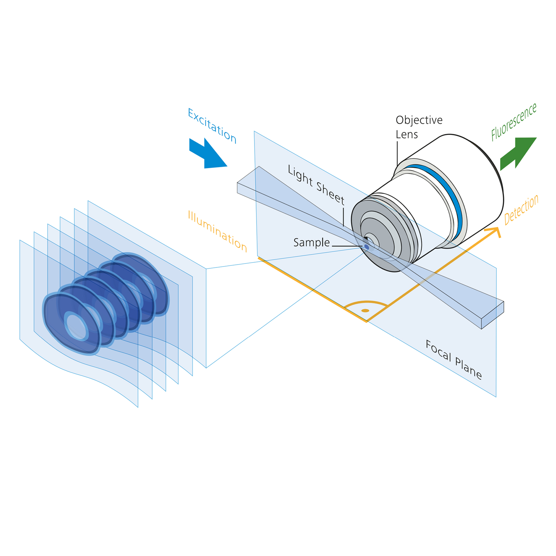
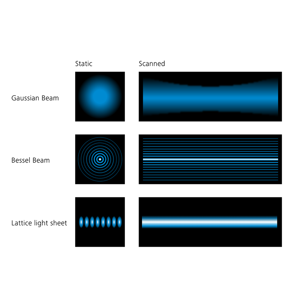
Conventional (Gaussian) light sheet microscopy splits fluorescence excitation and detection into two separate light paths, allowing to generate an inherent optical section by exciting only fluorescence from the in-focus plane.

Conventional (Gaussian) light sheet microscopy splits fluorescence excitation and detection into two separate light paths, allowing to generate an inherent optical section by exciting only fluorescence from the in-focus plane.

Light sheet microscopy

In general (also called Gaussian light sheet microscopy) is well known for its gentle imaging conditions at superior imaging speed. The groundbreaking concept of decoupling excitation and detection allows illumination of only the part of the specimen that is in the focal plane of the detection objective lens. By moving the sheet with respect to the sample and recording one image per focal plane, you can acquire volumetric data without exposing the out-of-focus sample areas.

Lattice Light Sheet Microscopy Principle

Lattice light sheet microscopy overcomes the limitations of Gaussian beams (limited optical sectioning, limited field of view) and Bessel beams (strong rings, excitation of out-of-focus fluorescence) by generating long and thin light sheets to achieve subcellular resolution.

Lattice light sheet microscopy overcomes the limitations of Gaussian beams and Bessel beams by generating long and thin light sheets to achieve subcellular resolution.

Lattice light sheet microscopy

Combines the advantages of light sheet microscopy with near-isotropic resolution in the confocal range. Advanced beam shaping technology creates lattice-shaped light sheets that are significantly thinner than standard Gaussian light sheets and thus provide increased resolution at comparable imaging speeds. The lattice structure of the light sheet is created using a Spatial Light Modulator (SLM), then projected onto the sample after passing scanners that dither the lattice structure to create a smooth light sheet.

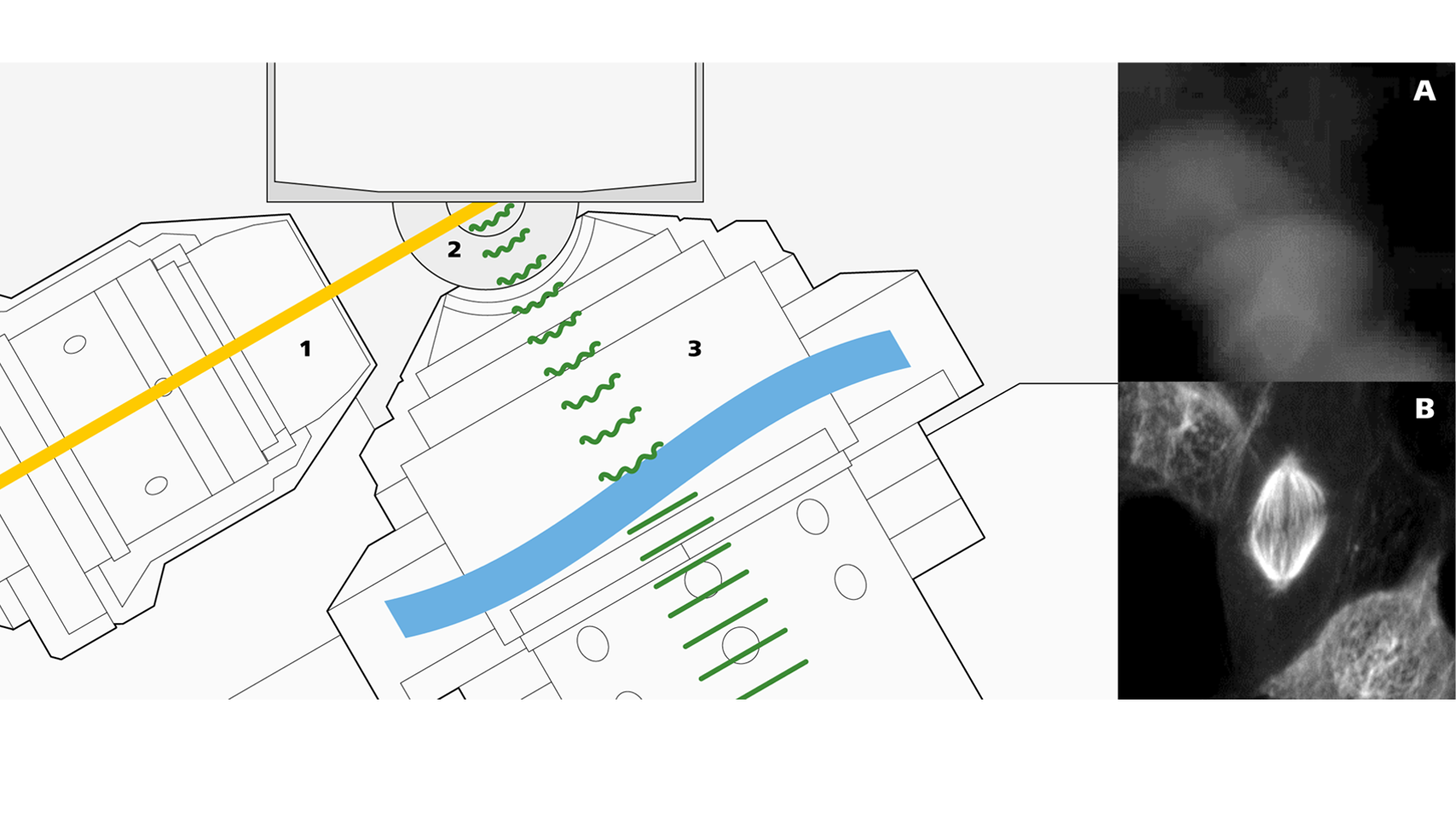
Schematic of sample carrier and core optics module with excitation objective (1), meniscus lens (2) and detection objective with free-form optics (3). Examples show imaging without (A) and with correction of refractive index changes (B).

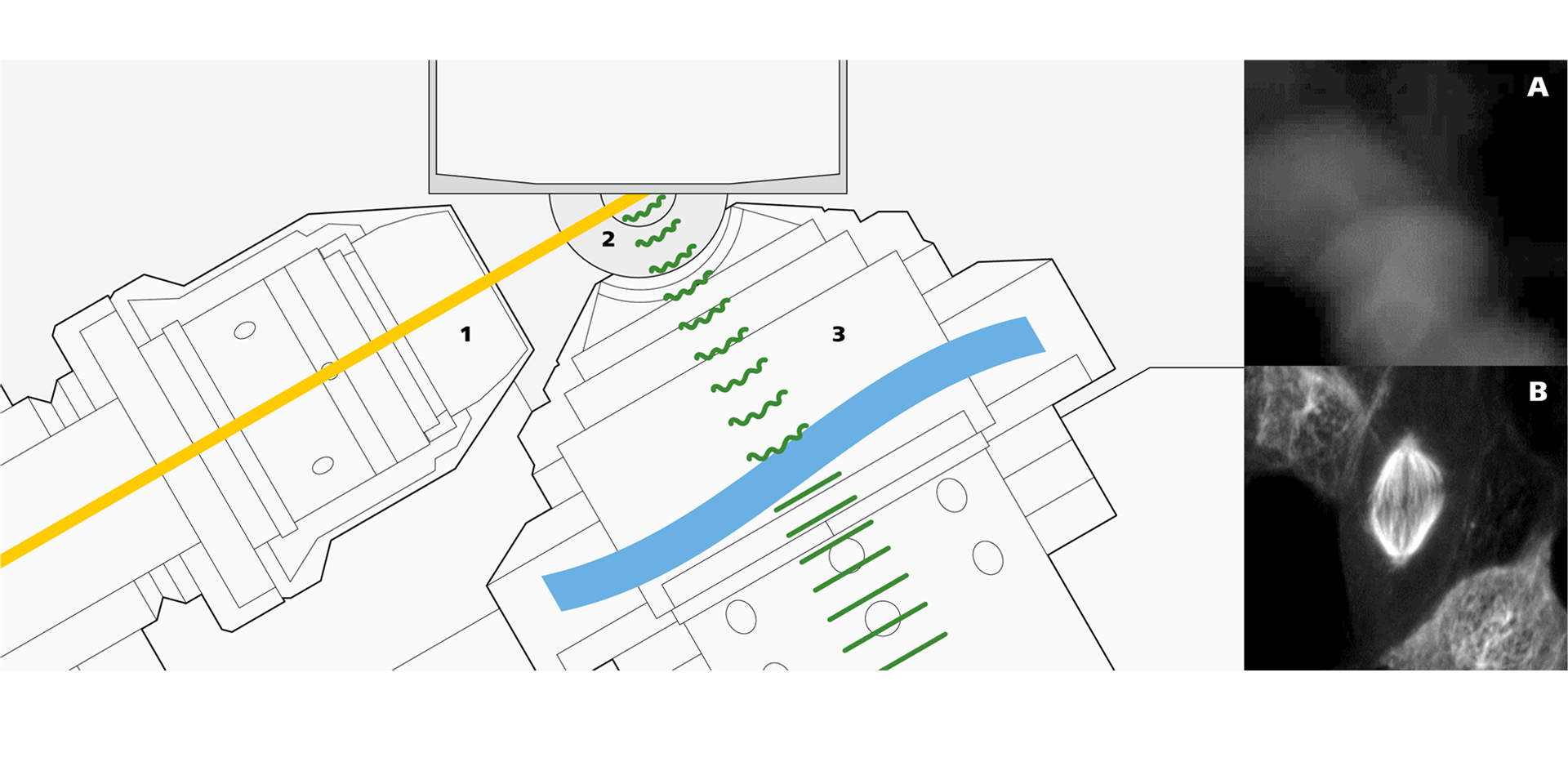
Schematic of sample carrier and core optics module with excitation objective (1), meniscus lens (2) and detection objective with free-form optics (3). Examples show imaging without (A) and with correction of refractive index changes (B).

Schematic of sample carrier and core optics module with excitation objective (1), meniscus lens (2) and detection objective with free-form optics (3). Examples show imaging without (A) and with correction of refractive index changes (B).

The ZEISS implementation of Lattice Light Sheet Microscopy

During the development of Lattice Lightsheet 7, ZEISS gave special attention to user-friendliness and compatibility with conventional sample preparation techniques. An inverse configuration is the most important prerequisite to allow the use of standard sample carriers for high-resolution microscopy. The challenges resulting from an inverse configuration are mainly refractive index mismatches as fluorescence is emitted from the sample, passes through aqueous cell culture media, a tilted glass coverslip and water immersion, then into the detection objective.

Unrivaled ZEISS optics

Special ZEISS proprietary optical elements in the detection beam path compensate for refractive index mismatches and enable you to image samples as easily and quickly as with a confocal microscope.

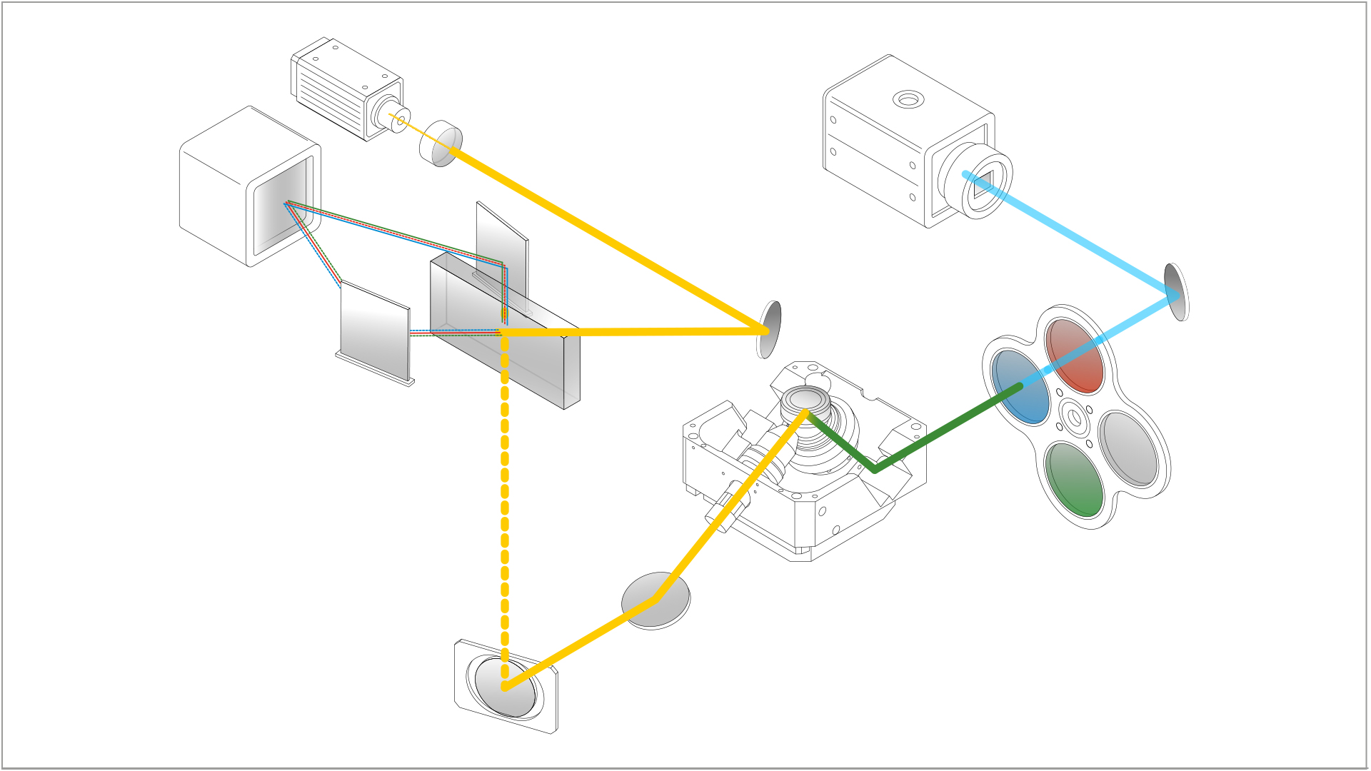
Schematic of the ZEISS Lattice Lightsheet 7 beam path

ZEISS Lattice Lightsheet 7 - Beam Path

Schematic of the ZEISS Lattice Lightsheet 7 beam path

The innovative design of the excitation beam path al lows for rapidly changing laser lines without having to reprogram the SLM. This enables virtually simultaneous acquisition of multi-channel data sets so that you will not miss any events occurring in your sample.

Schematic of the ZEISS Lattice Lightsheet 7 beam path (click to enlarge).

Automatic alignment of all optical elements

For the best imaging results, the lattice light sheet must be adapted to each sample; therefore, ZEISS has implemented automatic alignment of all optical elements to eliminate time-consuming manual adjustments. The innovative design of the excitation beam path allows for rapidly changing laser lines without having to reprogram the SLM. This enables virtually simultaneous acquisition of multi-channel data sets so that you will not miss any events occurring in your sample.

Downloads

    • ZEISS Lattice Lightsheet 7

      Long-term Volumetric Imaging of Living Cells

      12 MB


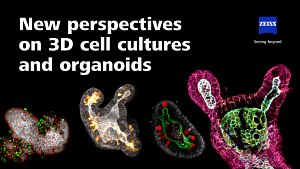
    • New perspectives on 3D cell cultures and organoids

      16 MB


Visit the ZEISS Download Center for available translations and further manuals.

Contact ZEISS Microscopy

Contact

Form is loading...

/ 4
Next Step:
  • Step 1
  • Step 2
  • Step 3
Contact us
Required Information
Optional Information

If you want to have more information on data processing at ZEISS please refer to our data privacy notice.