
Microdentistry in Endodontics
See more – treat moreApplication benefits of Microdentistry in Endodontics
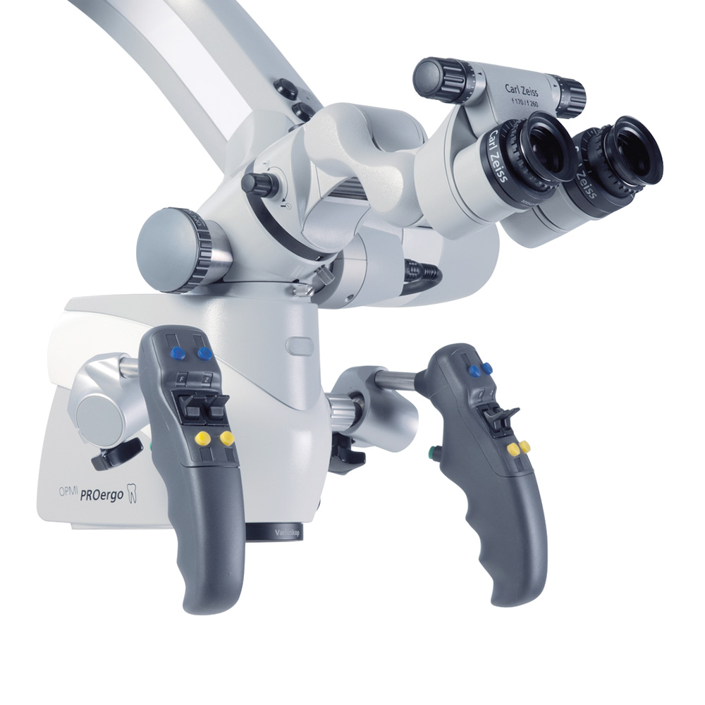
In the past root canal treatment was performed predominantly by feel. Visualization tools such as OPMI® from ZEISS allow you to view details and fine structures that are otherwise difficult to see with the naked eyes. Treatments can be performed with far greater precision and predictability than ever before – at any step of the endodontic workflow:


1 Canal location
Locate difficult-to-see anatomy such as calcified and accessory canals.


2 Cleaning & obturation
Control cleaning and obturation of root canals in all dimensions.


3 Examination of external tooth surface
Identify cracks, root fractures and external root resorptions.


4 Preservation of tooth structure
Remove tooth tissue during preparation and refinement of the access cavity strategically.
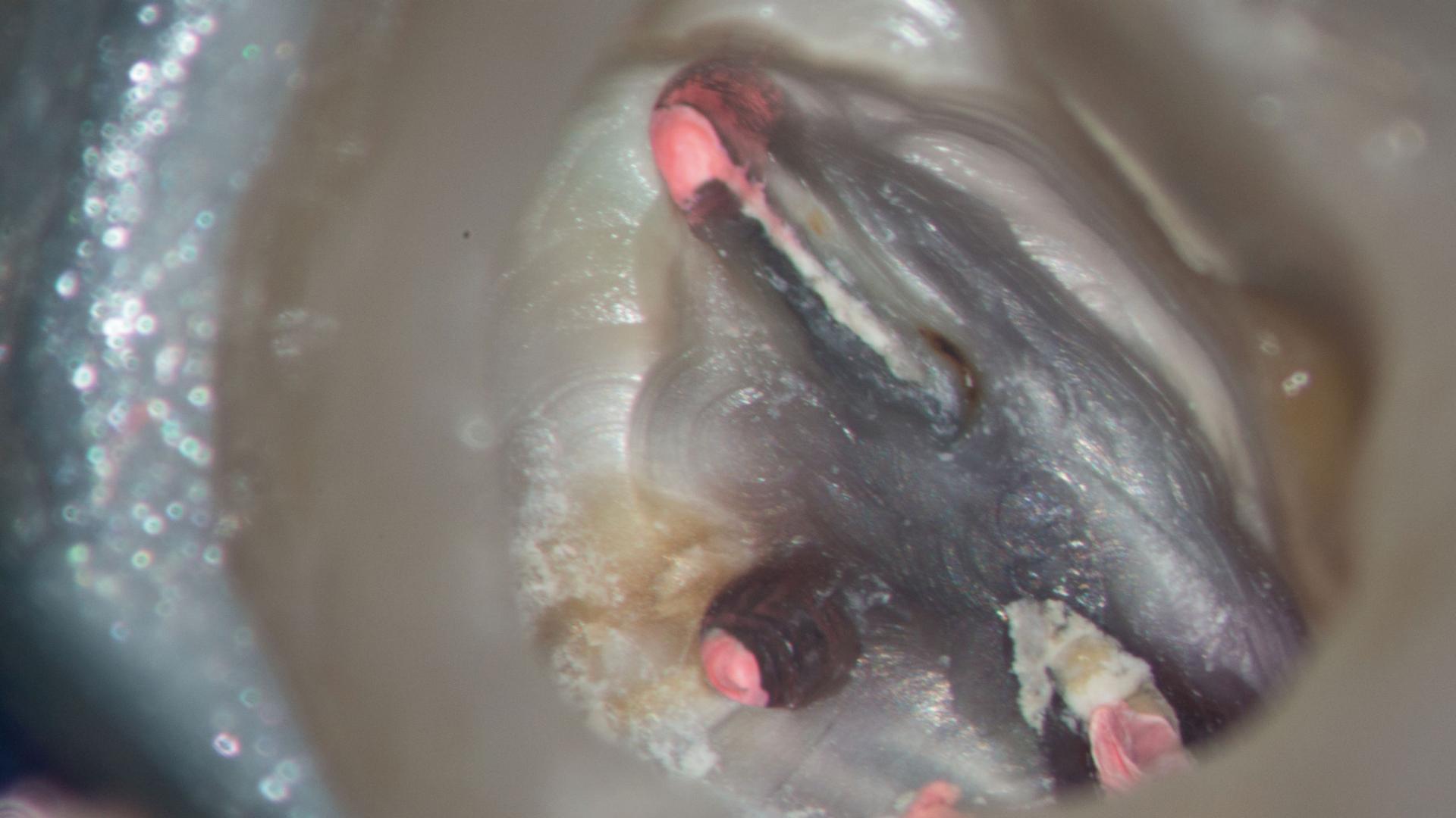
Canal location & cleaning
One of the greatest challenges in endodontics is locating canals, especially calcified canals. Canals sclerose from coronal to apical and several millimetres of sclerotic dentine may have to be removed before the canal is found. ZEISS OPMI tremendously facilitates this important part of endodontic treatment. Use medium to high magnification and maximum illumination when searching for small canals.
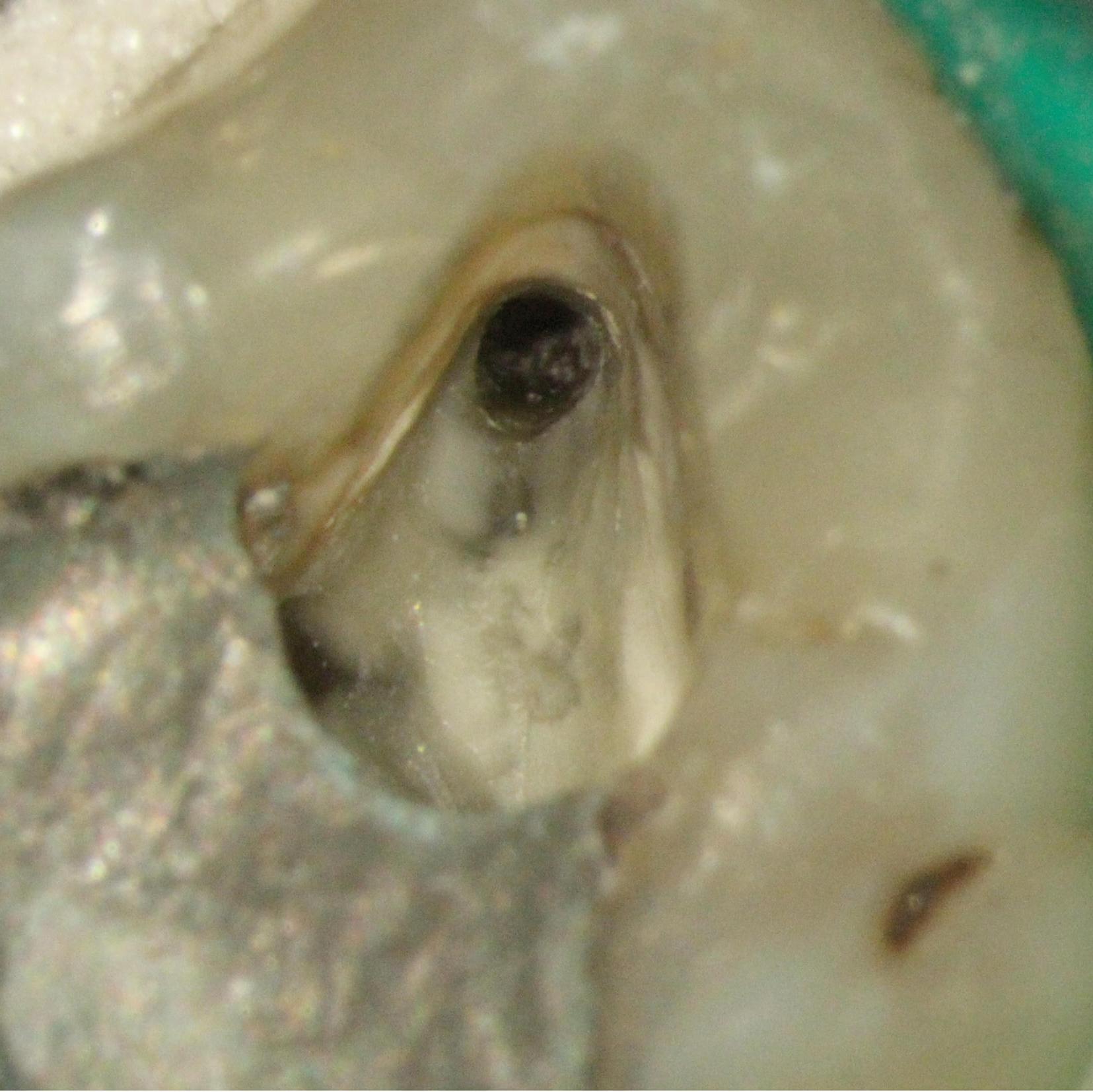

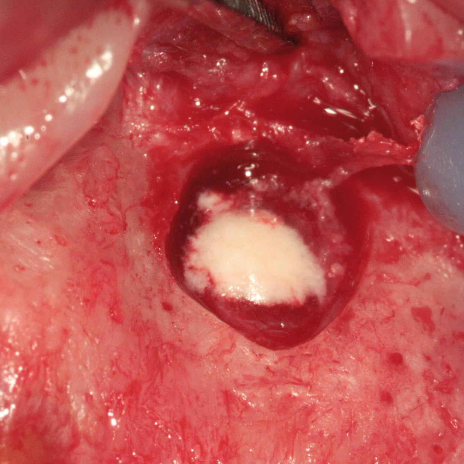
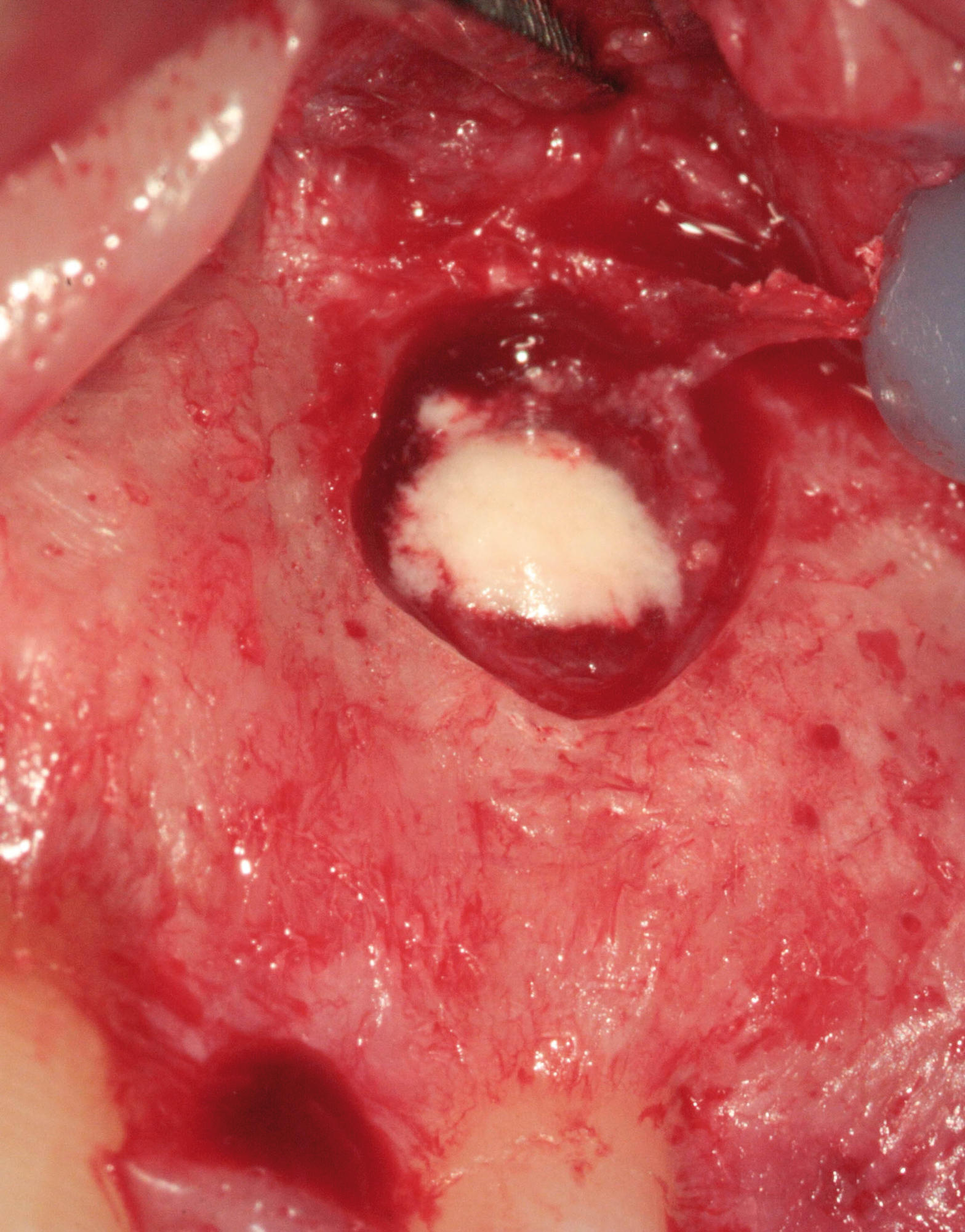
MB2 visible as a white spot, too small for a 0.6 file to enter.
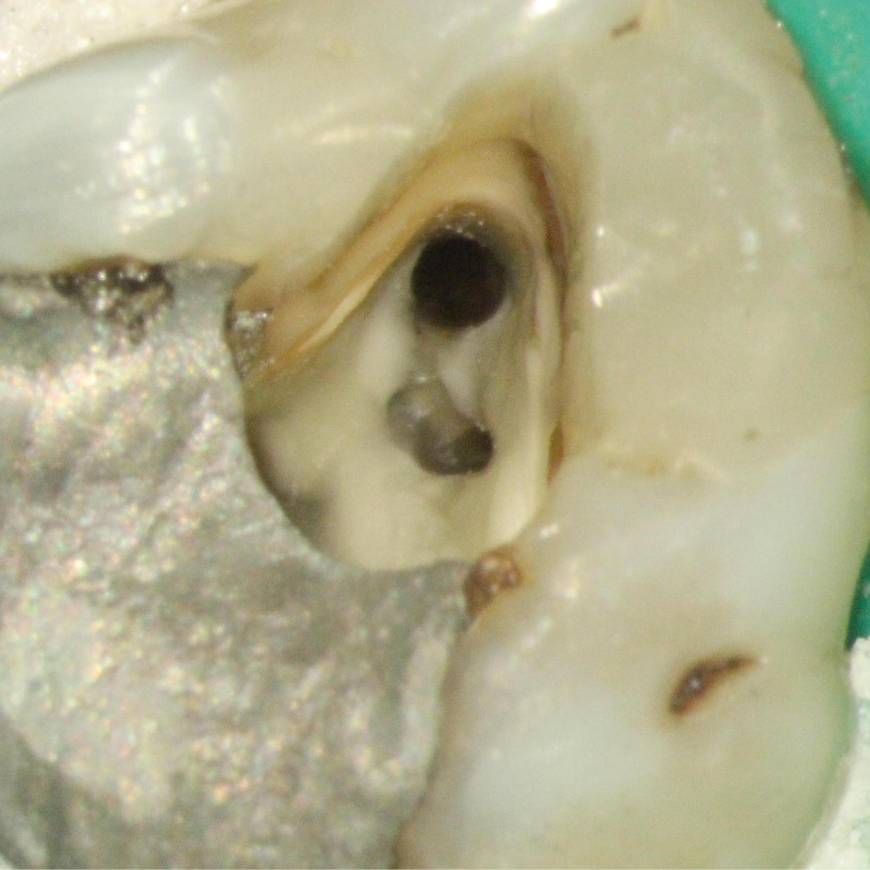


MB2 canal has been chased with a 0.5 mm rosehead bur and prepared with rotary files.
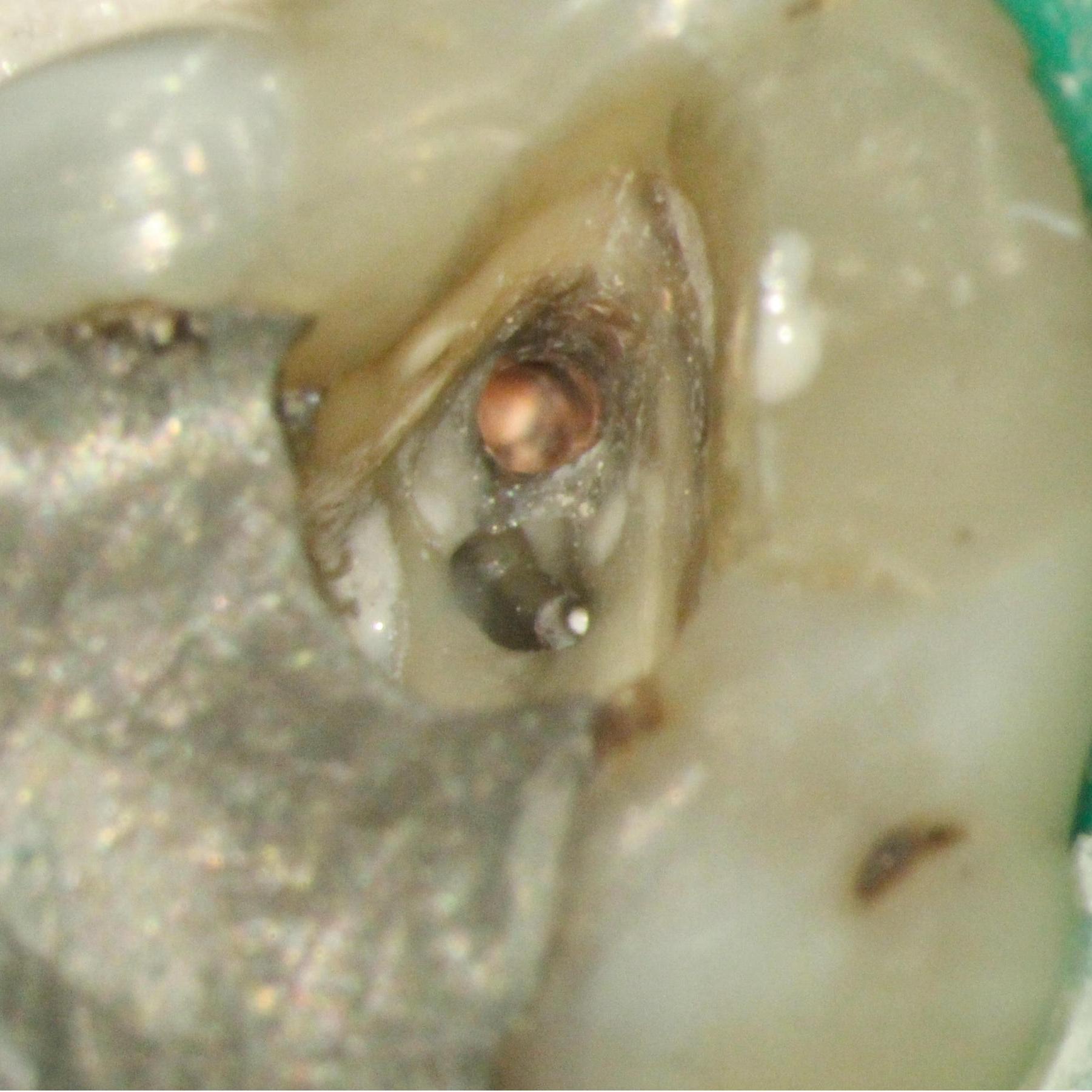

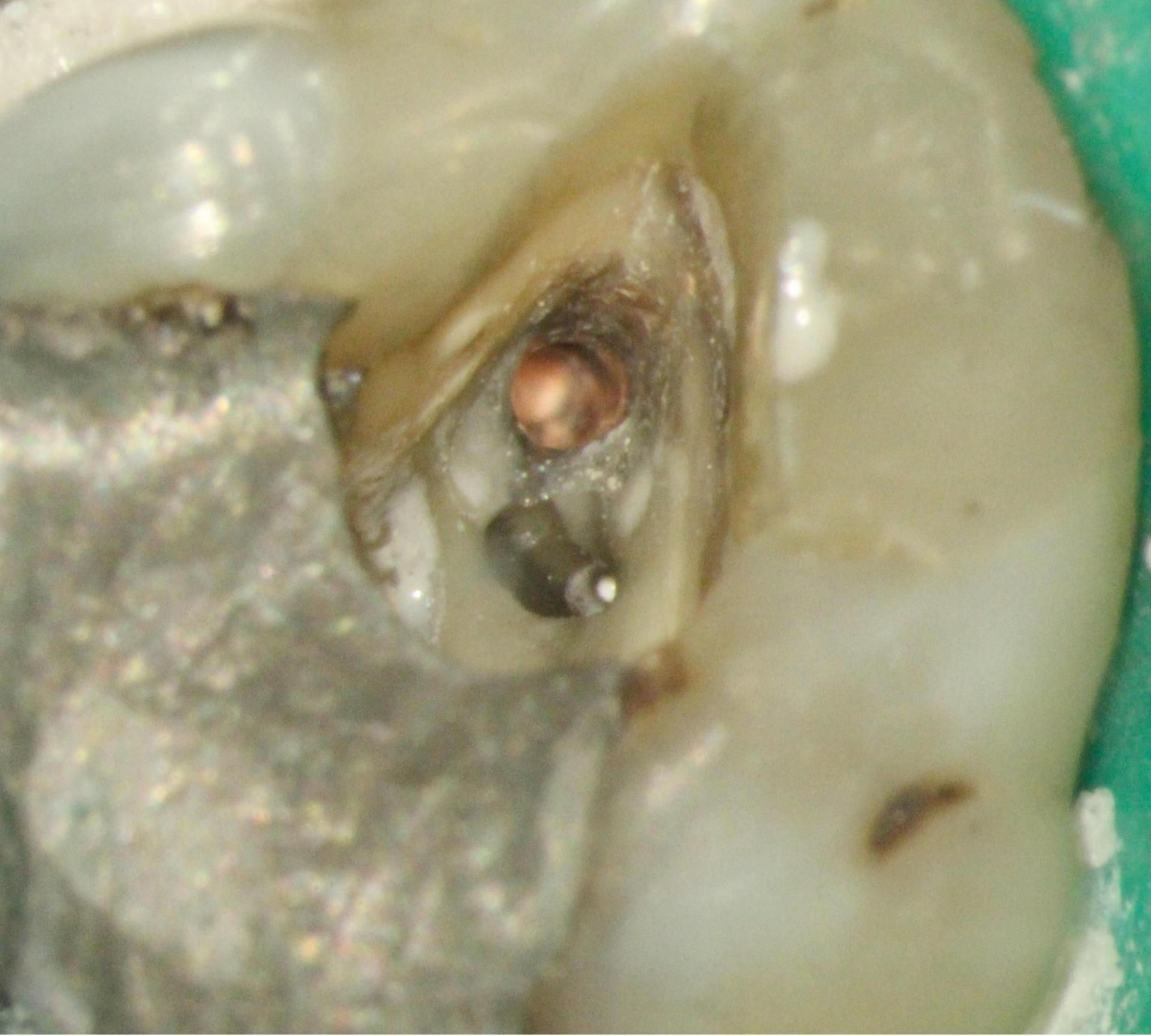

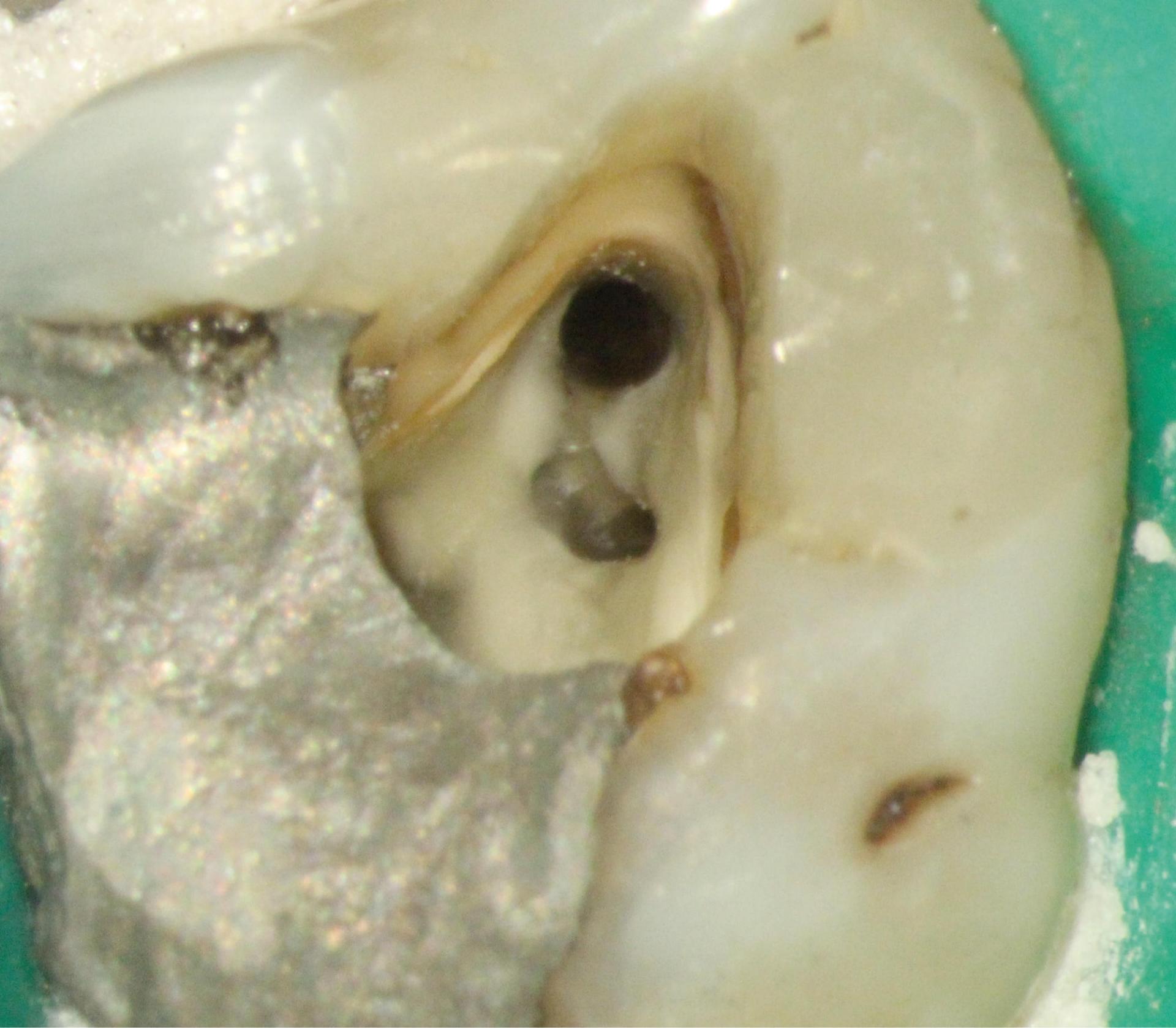
MB1 has been obturated and sealer has been forced into the MB2 canal indicating that MB1 and MB2.
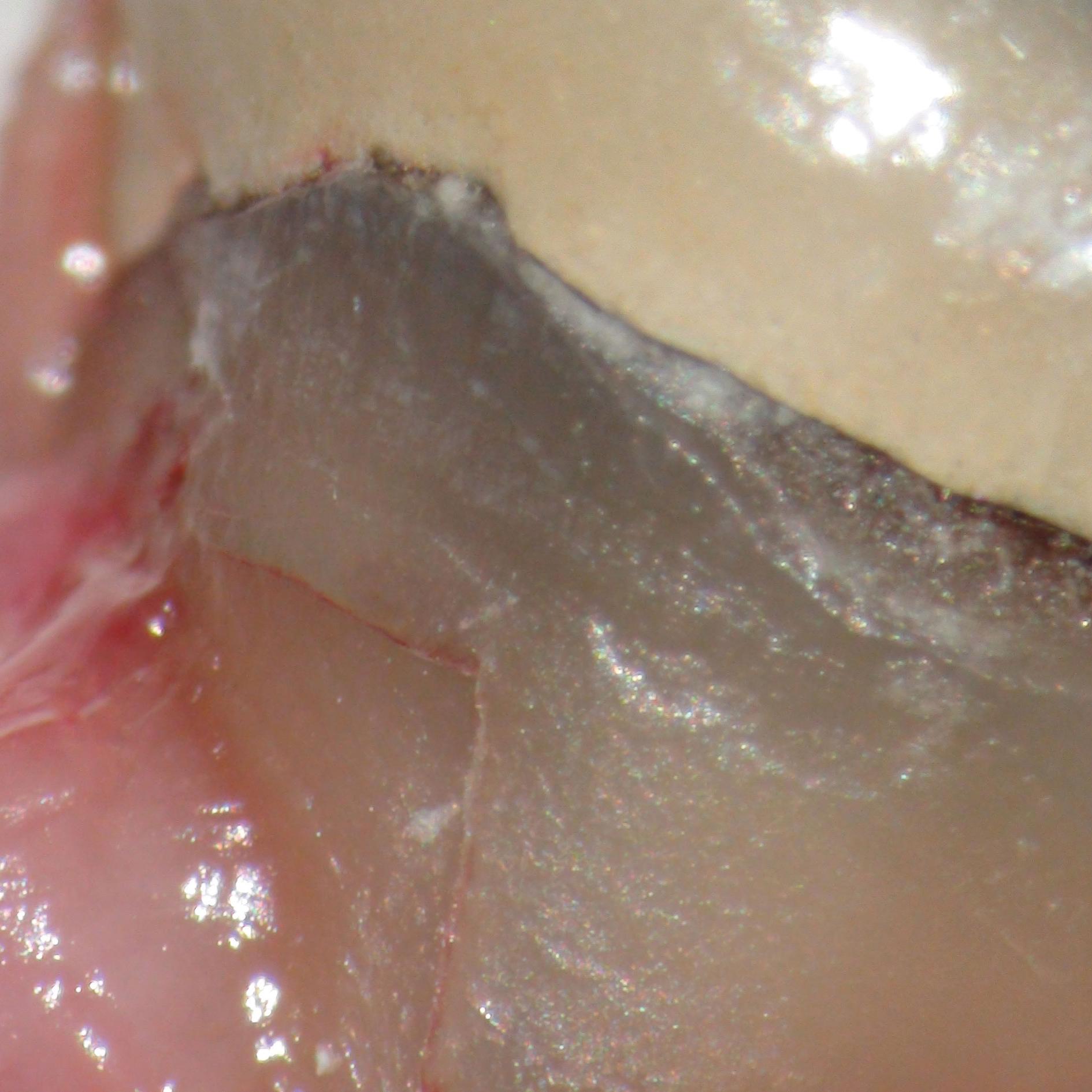
Endodontic Apicectomy
Thanks to the improved visualization provided by OPMI along with microsurgical instruments, this procedure can be performed much more conservatively. For instance, the amount of apical bone removal/osteotomy size does not need to be large when using OPMI. Hence, the procedure could be considered as minimally invasive.
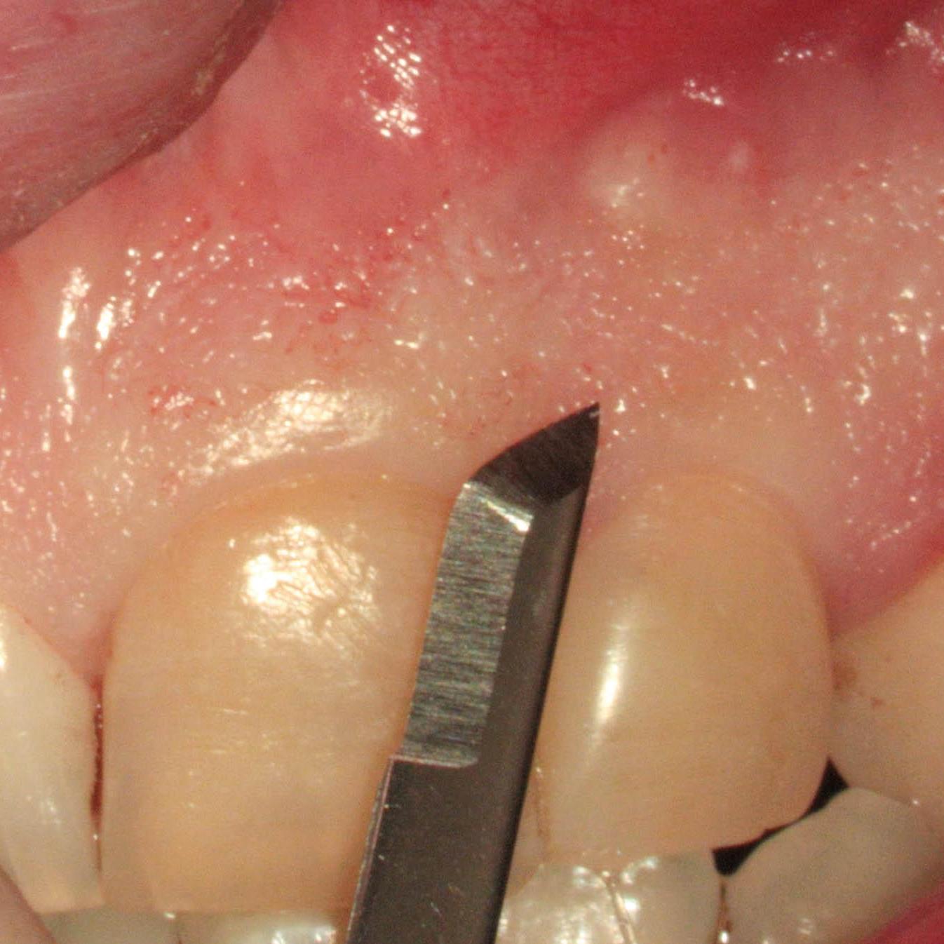

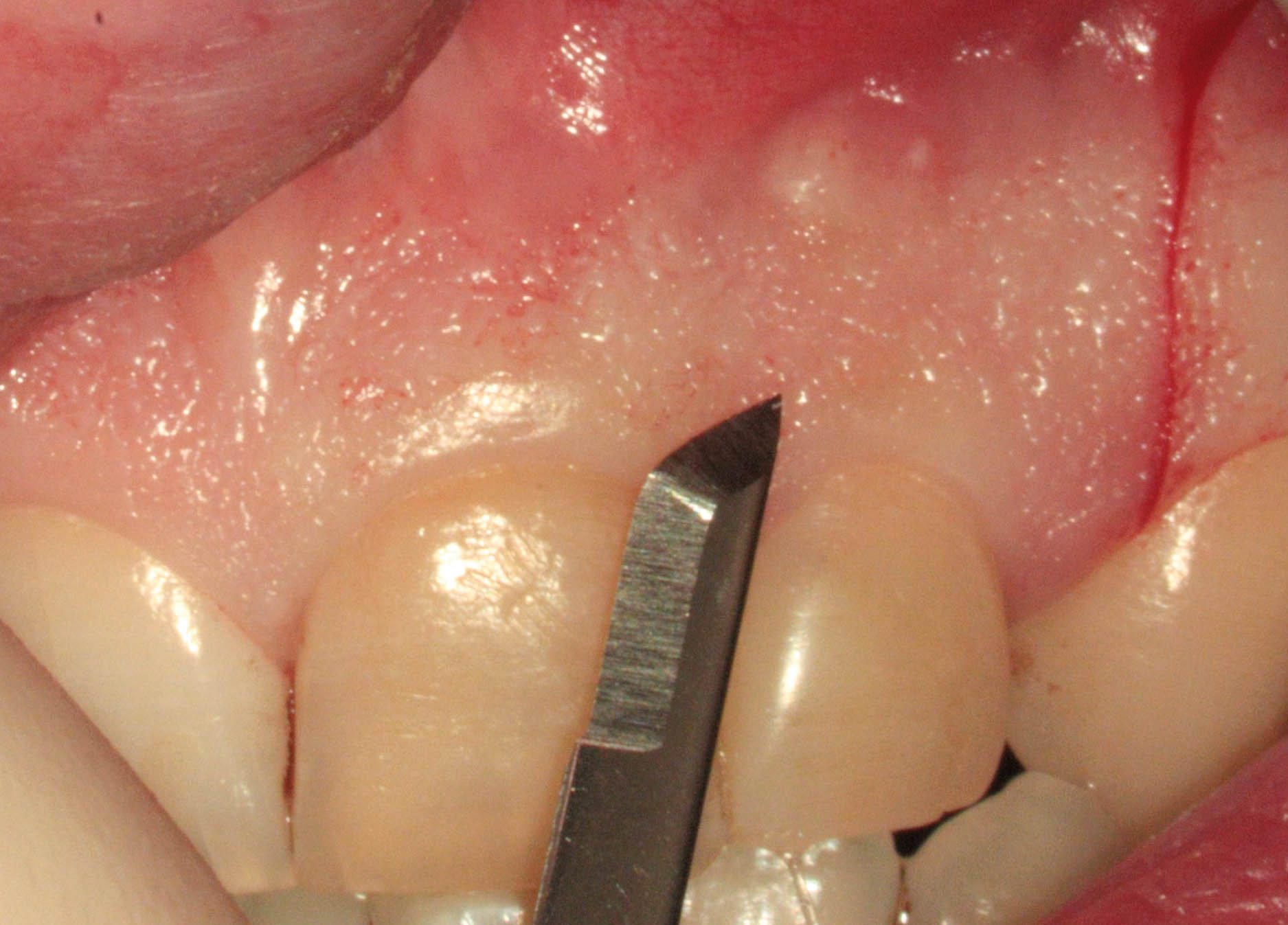

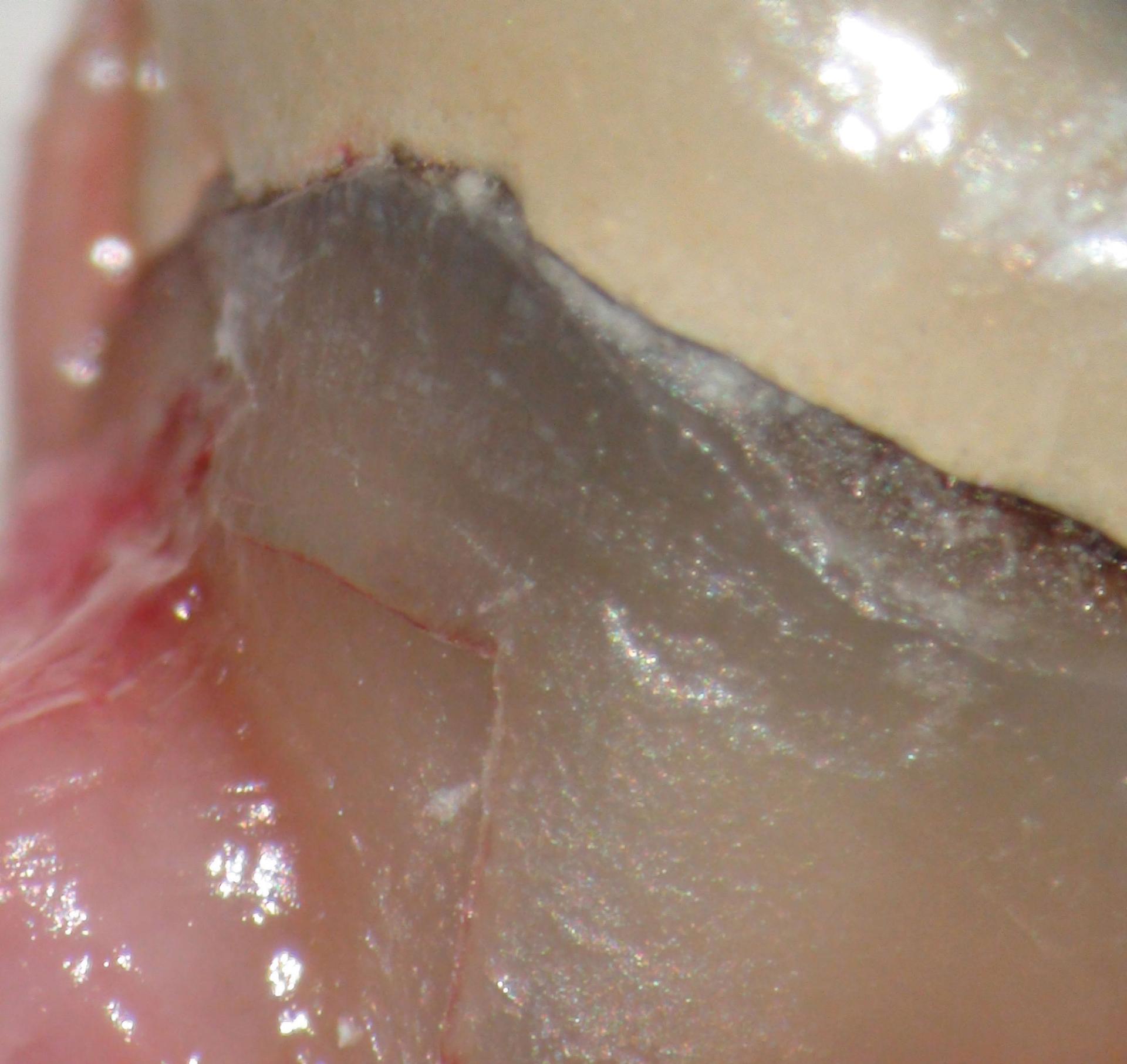
Fistula in 22 area; Micro-scalpel for atraumatic incision with tissue preservation.


Purulent discharge.
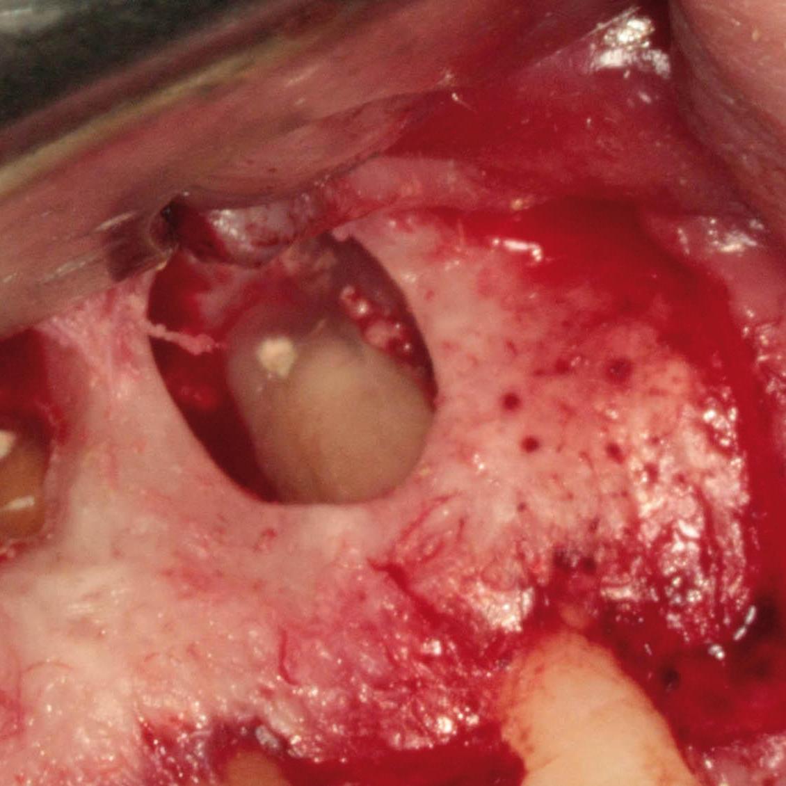

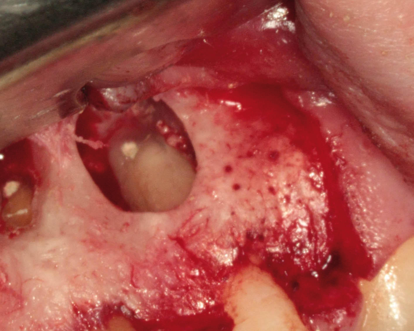

Obturation of the root chanel.
The root canal anatomy of teeth can be very variable and missed canals are a major cause of failure in root canal treatment. The OPMI tremendously facilitates this important part of endodontic treatment. It plays a vital role in helping to identify accessory canals at whatever level they may be.
Thanks to the improved visualization provided by the OPMI along with microsurgical instruments, endodontic root-end surgery can be performed much more conservatively. The procedure could be considered minimally invasive.
Endodontic re-treatment is considered to be one of the most challenging procedures in endodontics. In these situations the OPMI is essential.