Whether you have been in practice for one year or 35 years, the quest for improving efficiency and workflows is always top of mind for doctors. It is better for patients, better for your staff, and subsequently better for your practice. This is especially critical for small busy private practices like mine, where I work in tandem with only one optometrist. Consequently, it is critical that we work as efficiently as possible when it comes to providing care and leveraging the data we collect through examination and diagnostic testing to make clinical decisions.

The Evolution of Visual Field Testing
I have been in private practice for 35 years, taking over from my father who established our office in 1957. As a result, I have many glaucoma patients that we have followed for many decades. I still have some of our patients’ old charts where you can see the whole evolution of visual field testing. When I was a resident, I used to do the visual fields manually for my father, but then he got the first visual field machine that was available. At that time, visual field testing meant that either your patient saw the light or they didn’t. That was it. The test resulted in a printed-out punch card and did not provide any sort of comparison to previous testing. It was a suprathreshold test, so, unfortunately, by the time you were able to detect glaucoma, it was very advanced.
Since then, I’ve had all three generations of the Humphrey Visual Field machine and they just get slicker as time goes on—and faster, as well. The advances made in visual field testing have tremendously helped the workflow in our practice.
With the way that the testing protocols have evolved, as with SITA FASTER, we don’t need to worry about losing a technician for long periods of time when they need to run a visual field. This has enabled us to change our workflow patterns, where we can run a visual field on the fly on a patient without disrupting the schedule. Visual field testing can be a major bottleneck when it comes to workflows in a small practice, and now being able to do testing in half the amount of time but retain that reliability has added a layer of efficiency to how we move glaucoma patients in and out of the practice.
Additionally, it has tremendously helped improve patient satisfaction as we all know how much patients hate taking visual fields and how difficult it can be for them to maintain that level of concentration for long periods of time, which can ultimately lead to poor results.
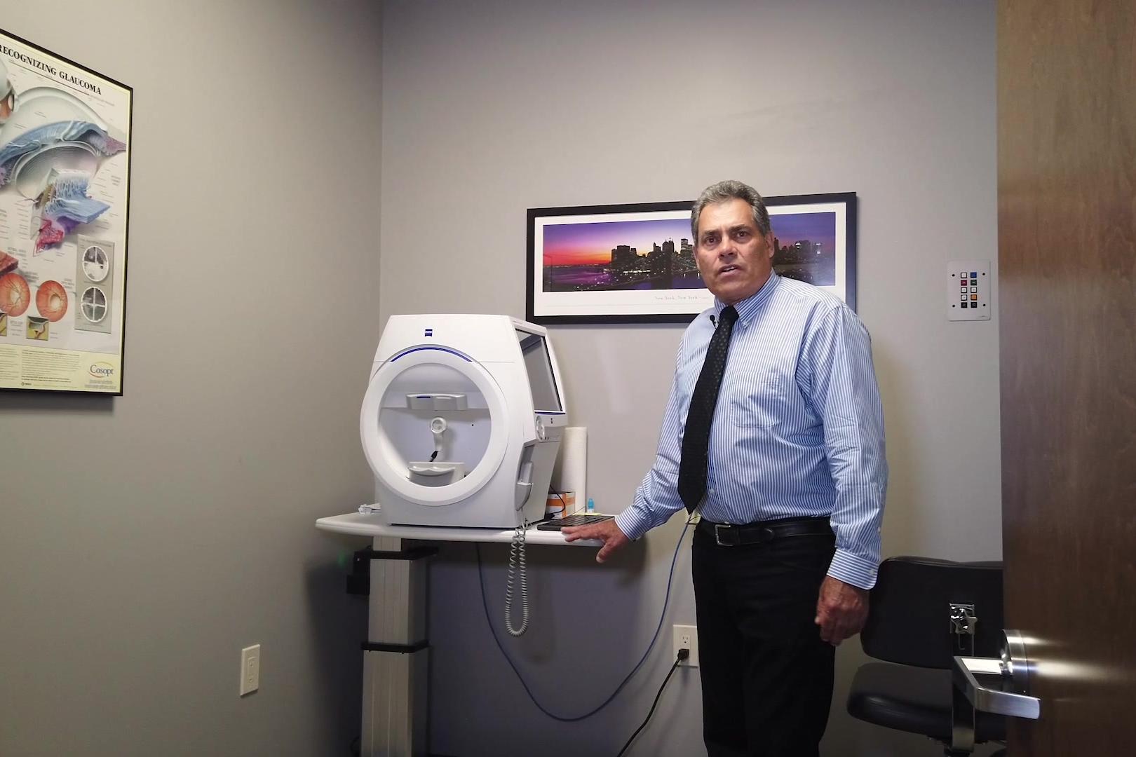
Dr. Douglas Liva: Efficient Workflow in Glaucoma Management
The Role of Optical Coherence Tomography (OCT)
Another major tool in our practice is obviously optical coherence tomography (OCT). OCT allows us to see changes in a way that visual fields do not, as structure versus function. It provides another level of data we can include in our analysis.
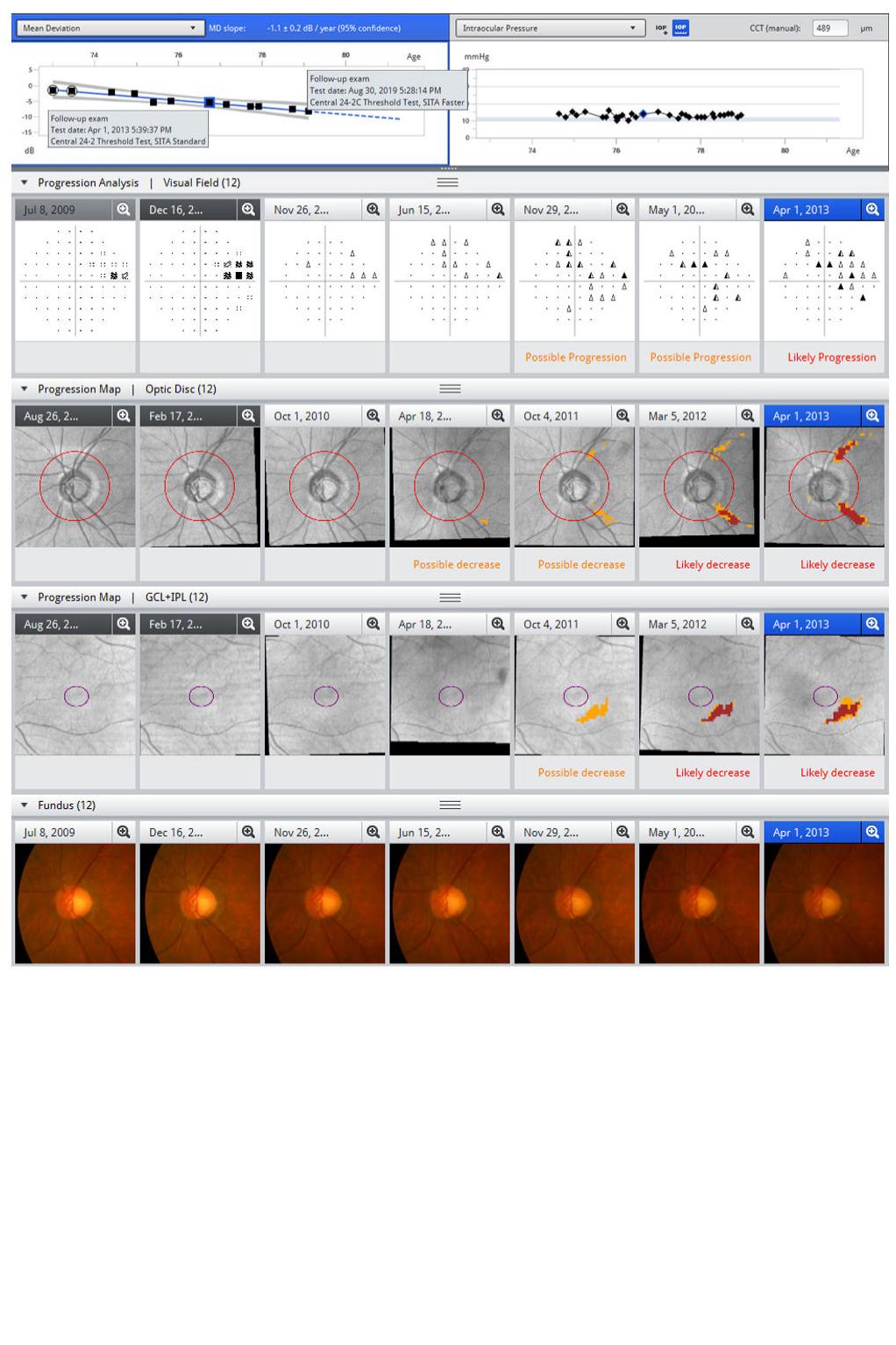
It also allows you to look at your visual field and compare it to your OCT. What are these tests telling you? Do they agree? Do they disagree?
Data comes down to numbers. You have the nerve fiber layer, the average thickness, the quadrants,
and the cup to disc ratio: all of this data is critically important in my workflow.
It presents a challenge, however, in that all these reports—whether you’re looking at a visual field, an OCT, or any other imaging—produces a series of numbers. What do you do with all of these numbers? And how do you handle all these numbers over years’ worth of testing?
Step one is capturing the data, and with visual field testing and OCT, we’ve been able to do that quite nicely. Step two, which is oftentimes the more challenging step, is taking that data and analyzing it
to make decisions.

The importance of being able to analyze data quickly and effectively
Capturing data is one thing, but analyzing it quickly, efficiently, and using it to make decisions is another. In a glaucoma practice, the importance of analyzing longitudinal data effectively is paramount.
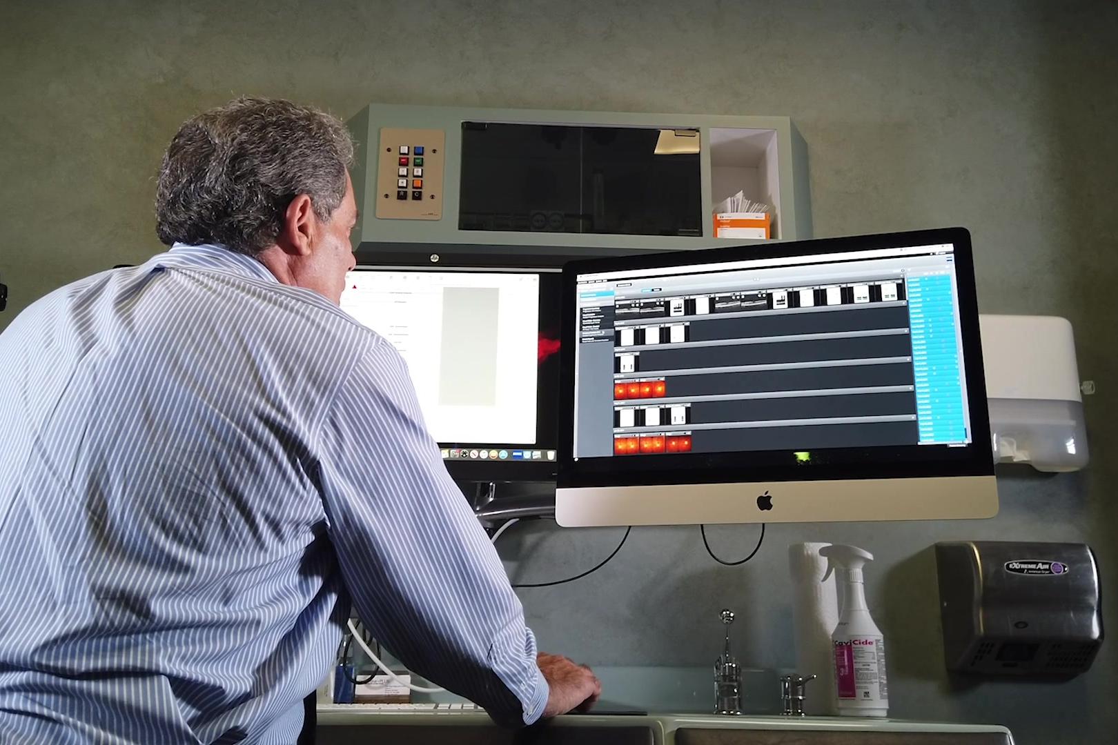
One of the most important tools we’ve adopted as a practice was FORUM.
Success in glaucoma management is not just about leveraging old data, but it is also about how you are incorporating and integrating new data. Between my optometrist and my three technicians, I am constantly being inundated with data. For a doctor accustomed to seeing six patients an hour, there is not enough time to spread out different visual fields or paper printouts on a table and try to evaluate the fields point by point.
With Glaucoma Workplace, I can crunch the data in literally seconds. It’s also more than just providing speed: it facilitates accuracy and ensures I am not missing things. It’s easy to miss little nuances, and that makes this tool especially important for those patients who I’ve seen over the years. On their OCT, their nerve fibers might still test normal and everything’s green; however, if you look at the progression analysis, there is a significant degradation.
How I Leverage Glaucoma Workplace
With Glaucoma Workplace, you can pull up data and results immediately. You can see if the cup has got bigger because it’s flagged with either red or yellow. You can see the difference in graphs as it’s colorcoded. It takes an endless amount of information across all of my diagnostic tools and tells me exactly what I need to know. I couldn’t live without it, frankly, and I can’t imagine practicing back where you had to manually figure things out.
I can only imagine how easy it is without Glaucoma Workplace to miss things because today, nobody can spend the proper amount of time. All the things that Glaucoma Workplace does, we could do progression analysis by printing tests and sitting down to study things, but who has that time?
However, just because Glaucoma Workplace allows me to spend less time with the patient, it does not mean I am diminishing the quality of my analysis. In fact, quite the contrary, and that is the true power of Glaucoma Workplace.
How FORUM has improved patient compliance
Not only does FORUM and Glaucoma Workplace drastically improve my workflow, efficiency, and clinical decision-making, it also has helped with patient compliance. In my office, I have two computer screens: one 27-inch screen that has my normal electronic medical records, and another 27-inch monitor with FORUM. Just like leveraging a fundus photo to show a patient where you’ve detected pathology, I can show the patient their progression.
If I can show them eight OCT results and each one is showing no difference, and then all of a sudden you see some yellow occurring, and then the yellow turns into red, it’s obvious that they’re progressing. So they know that something needs to be done. If I’m going to add another medication, or perform laser surgery or another intervention, it is far simpler to get that patient on board because they understand why I’m doing it.
I always explain to my patients exactly what my thought processes are. FORUM gives me a tool to explain better why.
Using this technology to be a non-treater
The decision to treat glaucoma is an important one: and just as important is the decision to not treat. Sometimes, deciding whether or not a patient has glaucoma, or predicting whether or not they will develop glaucoma can be extremely challenging. I prefer not to treat patients unless I have to, or unless I am confident they will develop glaucoma.
Today, the technology we have in the OCT and visual field—particularly with the contributions of SITA 24-2C—gives us the tools to make active decisions in patient care, even when that decision is to hold off on treatment. In order to be a non-treater, I need data to back up my decision. With in-depth longitudinal analysis, a thorough investigation into previous testing for comparison, and the addition of the SITA 24-2C which checks the central 10 degrees even more thoroughly than the normal 24-2, I feel far more confident about making these decisions.
So now, if I have a patient with higher pressures, but the OCT doesn’t budge and the visual fields are normal, I don’t feel any compulsion to treat them. And so I tell them, maybe I will have to treat them in the future, but every year that you don’t have to treat the patient is another year the patient doesn’t have the medication or treatment expense, the nuisance, and the side effects of medications.

The impact this has on my practice
Making the investment in these tools and in this technology pays dividends in so many ways. First and foremost, it improves decision-making, confidence, and the care I can provide to my patients. However, these tools not only impact the practice from a patient care perspective, but also from a practice growth perspective.
In terms of practice growth, you need to think about productivity. If you’re able to do the same job in less time, by definition, you are more productive. While I am performing procedures or examining patients, I have my technicians performing diagnostic testing, so that by the time I get into the exam room, Glaucoma Workplace is already loaded up. I walk in and everything’s right there and I can literally just make a decision and go over everything with the patient in a minute or two. Without these tools and technology, this would be very time-consuming, or I would have to take shortcuts. And when you take shortcuts, you make mistakes. This allows me to be more efficient without compromising quality.
The return doesn’t stop with productivity. It also impacts patient satisfaction, trust, and loyalty to our practice. When patients come to my office, they know they’re getting the best technology. Furthermore, if you are a patient and you have had your glaucoma managed by a doctor for 15 years who has been tracking all of your test results and analyzing data using a computer program, would you really want to go somewhere else and see another doctor? I don’t think so. Glaucoma is all about establishing a baseline and finding change. If you leave a practice and go elsewhere, even if I were to provide you with all of your testing printouts, you lose that raw data that tools like Glaucoma Workplace and FORUM allow you to retain.
When patients see what you offer, even if they did go somewhere else, especially a practice without technology for integrating the various test results longitudinally, everything would just look archaic and deficient. If this doctor started pulling out printouts or pulling up PDFs on screen, the patient will easily spot the difference.
Integrated diagnostic imaging is a powerful tool that can benefit your practice at every level, from diagnostics to patient compliance and satisfaction.