You are on our international English website. This site features our entire product portfolio worldwide. The products featured may not be available in the US. If you are a citizen from the US, please visit your country website for local information and contacts.
You are on our international English website. This site features our entire product portfolio worldwide. The products featured may not be available in the US. If you are a citizen from the US, please visit your country website for local information and contacts.
The video player is blocked due to your cookie preferences. To change the settings and play the video, please click the button below and consent to use of "Functional" tracking technologies.
Benefit overview
Third-party Content Blocked
The video player is blocked due to your cookie preferences. To change the settings and play the video, please click the button below and consent to use of "Functional" tracking technologies.
Experience of Dr. Sri Ganesh
Third-party Content Blocked
The video player is blocked due to your cookie preferences. To change the settings and play the video, please click the button below and consent to use of "Functional" tracking technologies.
Consistent visual outcomes1 enabled by ZEISS Optic (ZO)
Forgiving to decentration1
The sophisticated and patented ZEISS Optic (ZO) Asphericity Concept of the ZEISS CT LUCIA 621P/PY is designed to compensate for a wide range of aberrations due to different corneal shapes and lens positions. With its uniquely forgiving design it delivers excellent visual outcomes for a broad range of patients and surgical situations.
Picture showing that the IOL is not centered in the middle of the pupil.
Slit lamp examination showing that the pupil and the IOL are not aligned.
Does decentration matter?
Yes! Decentrations of varying magnitude are not uncommon. Besides the asymmetry of the eye, decentration of IOLs can occur due to poor capsular or zonular support, decentered capsulorhexis, asymmetric shrinkage of the capsular bag, misplacement of the haptics for IOL luxation in eyes with pseudoexfoliation. ZEISS CT LUCIA 621P/PY IOLs, with ZEISS optic features, are designed to compensate for potential decentration and lens misalignments. Reducing the risk of decentration allows you more time to focus on your patients and their needs.
The LUCIA lens has some special features […] you have the ZO Optic [ZO: ZEISS Optic], which compared to other aspheric lenses makes the lens more forgiving or less sensitive when it comes to decentration […] And therefore, we use this lens a lot, not only in challenging cases, when you expect, for example, phimosis or capsular shrinkage like pseudoexfoliation syndrome or after uveitis or traumatic cases, but also in standard cases.
Third-party Content Blocked
The video player is blocked due to your cookie preferences. To change the settings and play the video, please click the button below and consent to use of "Functional" tracking technologies.
Excellent stability
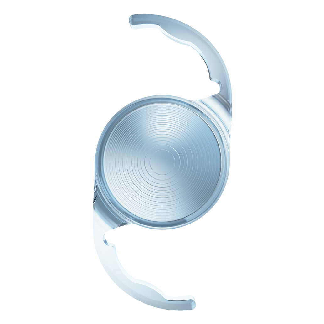
Maximized by direct capsular contact
ZEISS CT LUCIA 621P/PY IOLs feature an optic-haptic junction designed to ensure refractive stability. Coupled with step-vaulted C-loop haptics, this enables easy centering while maximizing direct capsular contact, thus ensuring stability and supporting a consistent, stable axial IOL position in the capsular bag.
The lens was very stable in the bag from the first week after surgery.
Third-party Content Blocked
The video player is blocked due to your cookie preferences. To change the settings and play the video, please click the button below and consent to use of "Functional" tracking technologies.
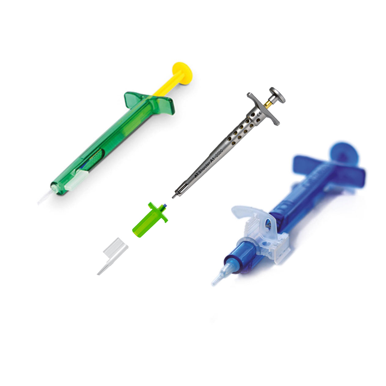
Intuitive injector handling
Easy to prepare. Smooth controlled injection
Third-party Content Blocked
The video player is blocked due to your cookie preferences. To change the settings and play the video, please click the button below and consent to use of "Functional" tracking technologies.
The video player is blocked due to your cookie preferences. To change the settings and play the video, please click the button below and consent to use of "Functional" tracking technologies.
Dr. Francisco Sánchez León, Mexico
Third-party Content Blocked
The video player is blocked due to your cookie preferences. To change the settings and play the video, please click the button below and consent to use of "Functional" tracking technologies.
Dr. Ishtiaque Anwar, Bangladesh
Third-party Content Blocked
The video player is blocked due to your cookie preferences. To change the settings and play the video, please click the button below and consent to use of "Functional" tracking technologies.
Dr. Sri Ganesh, India
Third-party Content Blocked
The video player is blocked due to your cookie preferences. To change the settings and play the video, please click the button below and consent to use of "Functional" tracking technologies.
Narayana Nathrayala Hospital, Bangalore
Dr. Naren Shetty, India
Third-party Content Blocked
The video player is blocked due to your cookie preferences. To change the settings and play the video, please click the button below and consent to use of "Functional" tracking technologies.
Italy
Dr. Antonino Cuttitta, Italy
Third-party Content Blocked
The video player is blocked due to your cookie preferences. To change the settings and play the video, please click the button below and consent to use of "Functional" tracking technologies.
Germany
Dr. Otmar Ringhofer, Germany
Third-party Content Blocked
The video player is blocked due to your cookie preferences. To change the settings and play the video, please click the button below and consent to use of "Functional" tracking technologies.
Sweden
Dr. Hossein Shams, Sweden
Third-party Content Blocked
The video player is blocked due to your cookie preferences. To change the settings and play the video, please click the button below and consent to use of "Functional" tracking technologies.
Austria
Dr. Andreas Borkenstein, Austria
I think the CT LUCIA 621 comes closest to the ideal intraocular lens.
Publications
Han et al. - CT LUCIA 621P vs TECNIS Eyhance ICB00 | Study Spotlight EN
Please note: ZEISS CT LUCIA 621P/PY and ZEISS CT LUCIA 611P/PY IOLs share the same hydrophobic acrylic material and very similar optic and lens designs. Therefore, the reported results in publications for CT LUCIA 611P/PY also apply to 621P/PY IOLs.
Product Brochure
CT LUCIA 221P / 621P / 621PY Compendium Interactive version with Live Links EN
The data is taken from a simulation. The transferability of the results of such a simulation to patients with an actual implanted intraocular lenses has not yet been scientifically proven. Whether the simulated impressions correspond to the actual visual impressions must be clarified in future invasive studies.