You are on our international English website. This site features our entire product portfolio worldwide. The products featured may not be available in the US. If you are a citizen from the US, please visit your country website for local information and contacts.
You are on our international English website. This site features our entire product portfolio worldwide. The products featured may not be available in the US. If you are a citizen from the US, please visit your country website for local information and contacts.
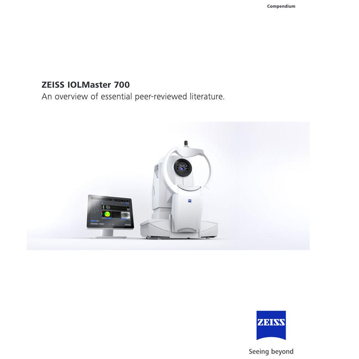
Thanks to SWEPT Source OCT technology, ZEISS IOLMaster 700 offers unique features and functions to achieve fewer refractive surprises.
Total Keratometry: replacing assumptions with measurements
The Total Keratometry (TK®) of ZEISS IOLMaster 700 allows you to directly measure the posterior corneal surface using SWEPT Source OCT. In a study the exclusive Barrett True K with TK formula improved the outcome prediction compared to the Barrett True K with Classic Ks within ±0.5 D by >12 % (p = 0.04) in post-myopic LASIK eyes1.
The Barrett True K with TK formula elevates post corneal refractive surgery IOL power calculation to the next level.
Third-party Content Blocked
The video player is blocked due to your cookie preferences. To change the settings and play the video, please click the button below and consent to use of "Functional" tracking technologies.
Unique Fixation Check
Can you see the foveal pit? If so, you can reduce the risk of refractive surprises due to incorrect measurements caused by undetected poor fixation.
Detect unusual eye geometries and visually verify your measurements
The patented Cornea-to-Retina Scan allows you to detect unusual eye geometries, such as tilt or decentration of the crystalline lens. Additionally, the complex interpretation of A-scans is no longer necessary.
Optimize your cataract workflow
With the ZEISS IOLMaster 700, you enjoy exceptional speed, an outstanding cataract penetration rate, and benefit from the unique, patented Fixation Check. Combined with the EQ Workplace® cataract surgery planning software from ZEISS, you can streamline your cataract workflow from the office to the OR and back and access your biometric data anytime, anywhere.
Third-party Content Blocked
The video player is blocked due to your cookie preferences. To change the settings and play the video, please click the button below and consent to use of "Functional" tracking technologies.
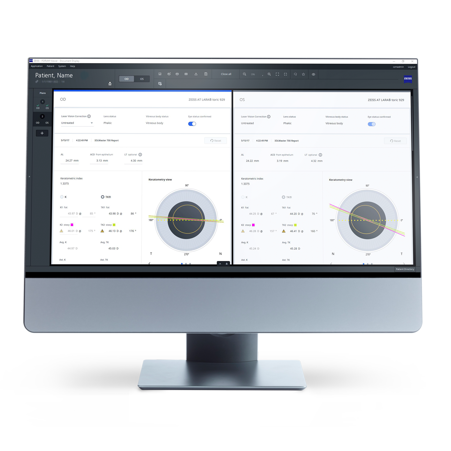
Connect to ZEISS EQ Workplace
Connect your ZEISS IOLMaster 700 with ZEISS EQ Workplace cataract surgery planning software. It supports your biometry data review and analysis, IOL calculation and selection, IOL ordering, surgical planning, and post-operative data collection.
Save time during pre-operative processes
Further protect against never-events
Access your data from anywhere
Personalize your IOL constants
Third-party Content Blocked
The video player is blocked due to your cookie preferences. To change the settings and play the video, please click the button below and consent to use of "Functional" tracking technologies.
Measurement in less than 45 seconds
With ZEISS IOLMaster 700, you can measure both eyes in less than 45 seconds. Alignment assistance functions make the results largely independent of the user and therefore easy to delegate.
Case with a very dense cataract that could be measured successfully with ZEISS IOLMaster 700. Image courtesy of Prof. M. de La Torre, DLT Ophthalmic Center, Peru.
Cataract penetration up to 99 %
A comparative clinical study with more than 1,200 eyes showed that ZEISS IOLMaster 700 achieves a cataract penetration rate of up to 99 %. As a result, the number of ultrasound cases can be reduced by 92 %, saving you valuable time.2
Macular hole: Fixation Check image (middle) combined with ZEISS CIRRUS retina OCT image (via photo editor program). Image courtesy of Prof. W. Sekundo, Phillips University Hospital Marburg, Germany.
Indications for macular pathologies with Fixation Check
Although ZEISS IOLMaster 700 is clearly not intended to be used for diagnostics3, in high-volume practices the ability to detect these eyes preoperatively can be invaluable.4
Central Topography to start your workflow with more insights
With no changes in workflow, the IOLMaster® 700 from ZEISS now measures Central Topography. It allows easy reading of central corneal shape information when you start your workflow and before you decide on the IOL and consult with your patient.
It is integrated into the standard biometry measurement of the ZEISS IOLMaster 700, without the need of any additional hardware or measurement time. The scaling and hues have been developed in cooperation with Douglas D. Koch, MD, and Li Wang, PhD, USA.
It is remarkable how much information we get from Central Topography.
See clinical evidence: Compendium of essential peer-reviewed literature
The ZEISS IOLMaster 700 could be just the beginning: Take your workflows to a new level and learn how our products and applications add value beyond the mere sum of their parts.
Lawless M, Jiang JY, Hodge C, Sutton G, Roberts TV, Barrett G. Total keratometry in intraocular lens power calculations in eyes with previous laser refractive surgery. Clin Exp Ophthalmol. 2020 Aug;48(6):749-756
2
R. Varsits, N. Hirnschall, B. Doeller, O. Findl; Increasing the number of successful axial eye length measurements using swept-source optical coherence tomography technology compared to conventional optical biometry; presented at ESCSR 2016.
3
Findings need to be verified and pathologies diagnosed with a dedicated retina OCT.
4
Hirnschall N, Leisser C, Radda S, Maedel S, Findl O. Macular disease detection with a swept source optical coherence tomography based biometry device in patients scheduled for cataract surgery. JCRS VOL 42, APRIL 2016. Bertelmann et al.; Foveal pit morphology evaluation during optical biometry measurements using a full-eye-length swept-source OCT scan biometer prototype; European Journal of Ophthalmology, Nov/Dec 2015.