
ZEISS PLEX Elite 9000 Swept-Source OCT Angiography
Uncovering the Undiscovered
PLEX® Elite 9000 from ZEISS is a transformational OCT imaging technology that invites retina and glaucoma researchers into a new world of structural and microvascular clarity of the anatomy.

The First Dual-Speed Swept Source OCT
Faster, deeper, and with higher resolution, the new dual-speed PLEX® Elite 90001 elevates ophthalmic imaging and clinical research to a new level. With this technology, clinicians can support longitudinal studies while expanding the scope of their research to investigate the clinical benefits of scans acquired at 200kHz scan speed.

Explore Deeper Meanings
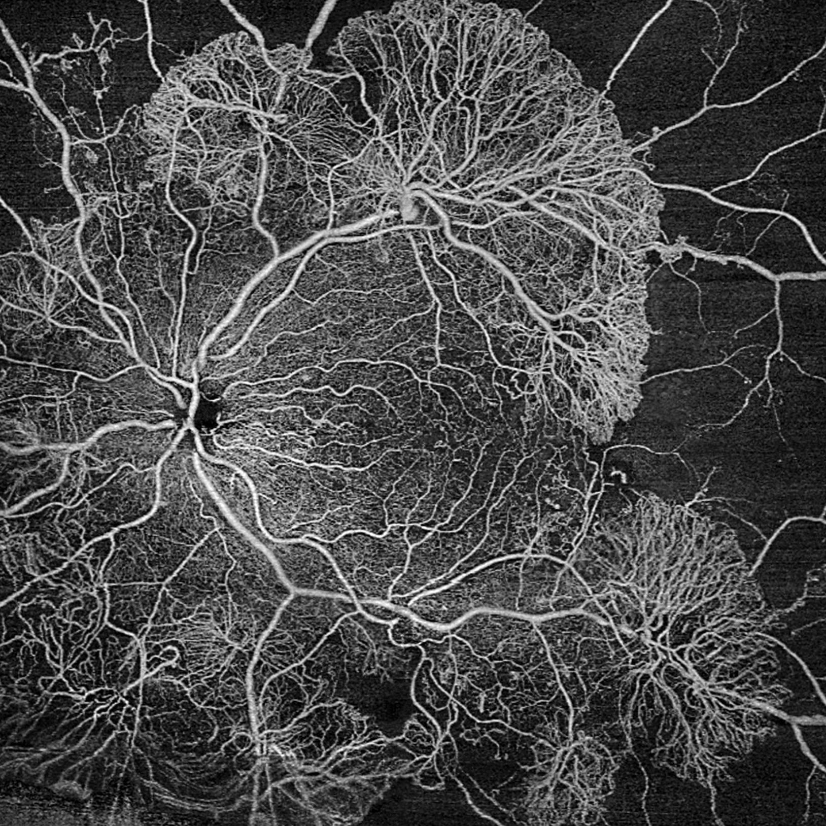
ZEISS PLEX Elite 9000 Swept-Source OCT allows clinical researchers the potential to see deeper, wider and in more detail from the vitreous to the sclera in the posterior segment.
- Study early mechanisms of micro- and neovascularization of the posterior segment from vitreous to sclera.
- Explore the progression of retinal and choroidal pathology, such as CNV
- Evaluate the mechanism of retina and choroid response to a therapy.
Pick a scan sequence that’s right for your case
- Ultra-wide 15x15 scan provides a large field of view captured in a single OCT angiography scan
- High-density OCT angiography scans reveal vasculature details in unparalleled clarity
- UHD spotlight high detail B-scan up to 16 mm width and 6 mm depth
- Montage capability to cover a combined field of view of up to 70 degrees
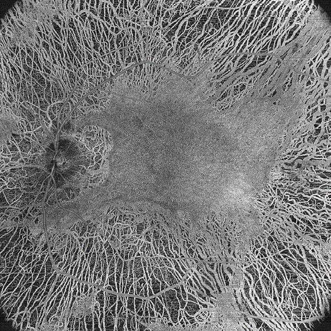

Widefield 15x15 mm Swept-Source OCT Angiography image of Retinitis Pigmentosa scanned at 200kHz scan speed.
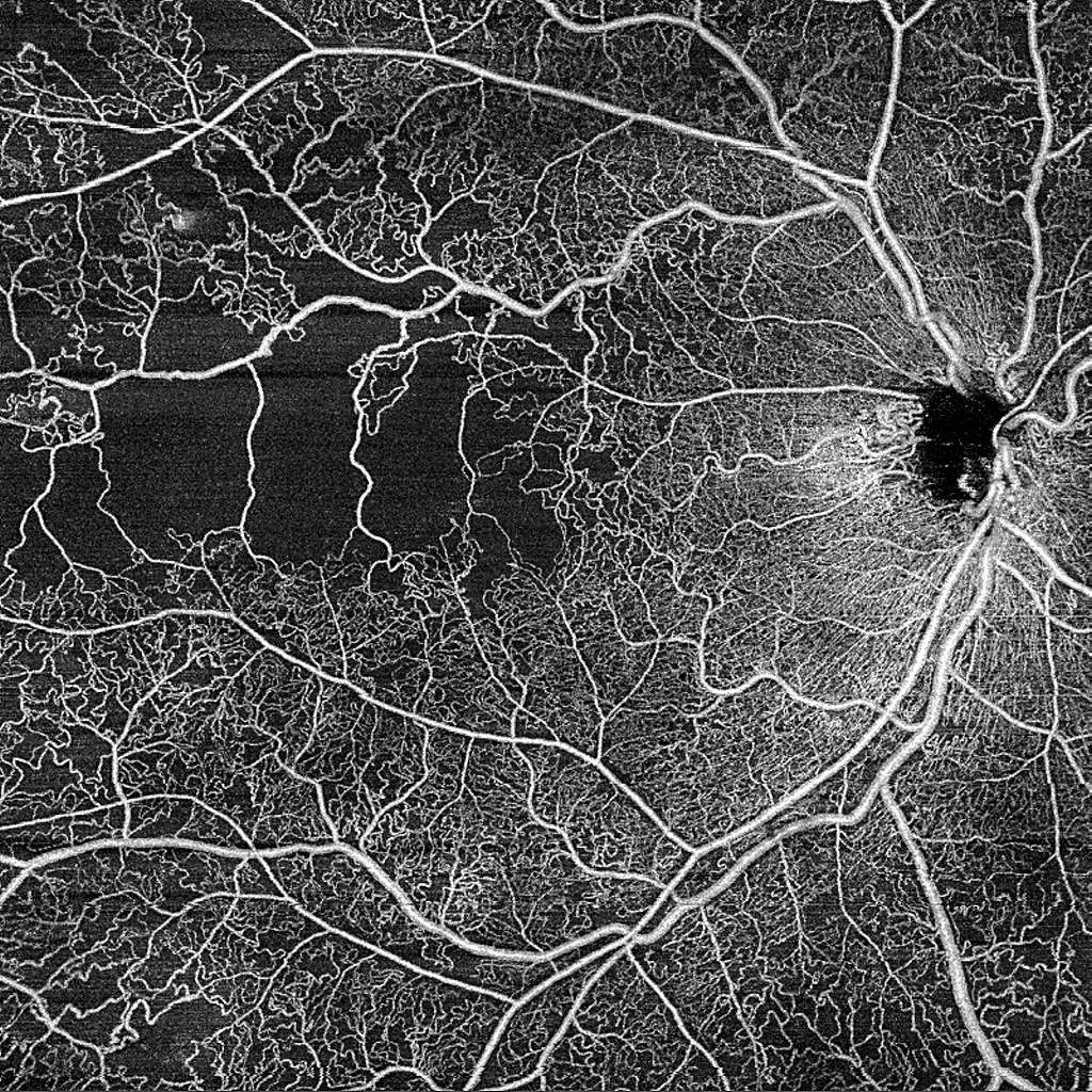

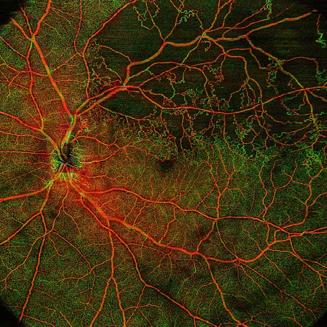
HD Angio 12x12mm Swept-Source OCT Angiography of Branch Retinal Vein Occlusion scanned at 200kHz scan speed

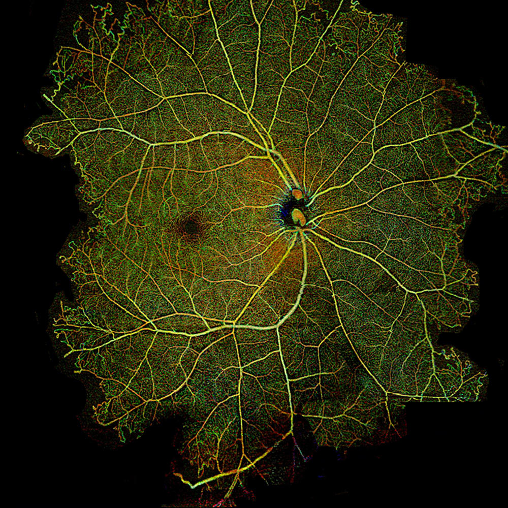
Widefield 15x9 AngioPlex map of a Choroidal Neovascular Membrane, RPE/RPE Fit layer
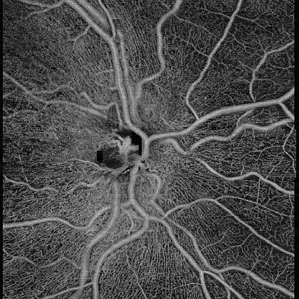

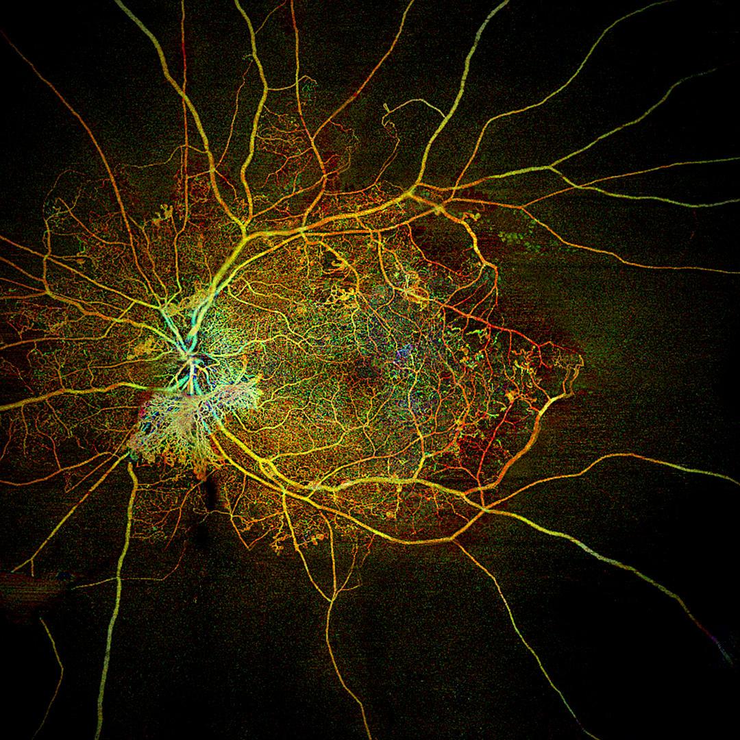
Angio 6x6 mm Swept-Source OCT scan of the Optical Nerve Head with Intracranial Hypertension
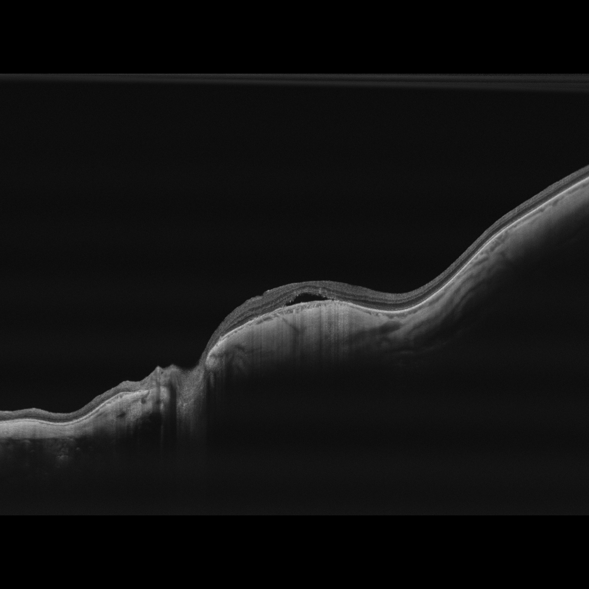

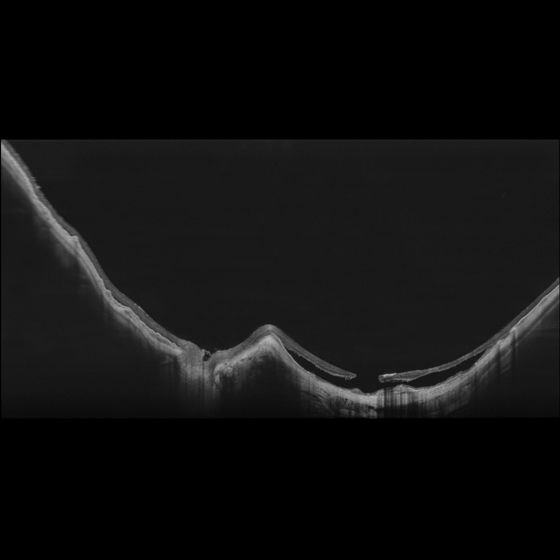
UHD Spotlight Swept-Source OCT B-scan image of Myopia, Serous Retinal Detachment and Dome-Shaped Macula with 6mm scan depth and higher-resolution scanned at 200kHz scan speed

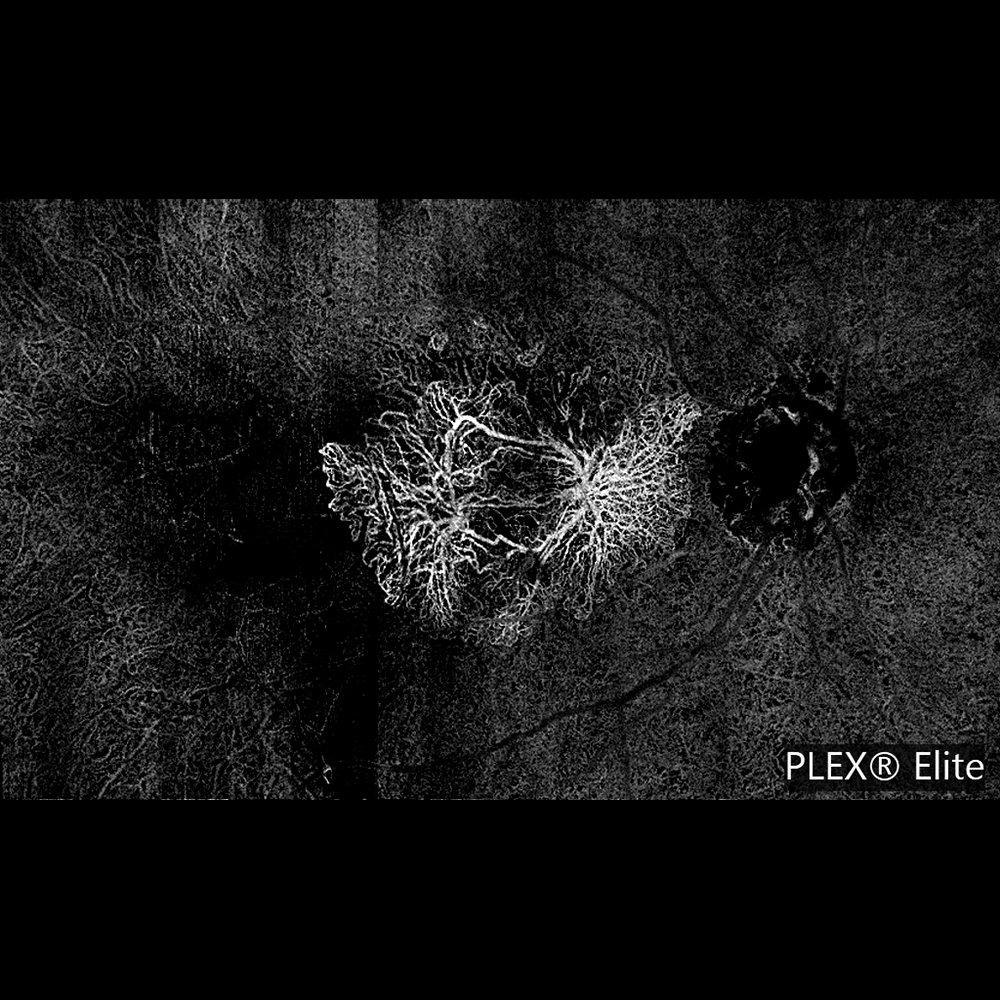
UHD Spotlight Swept-Source OCT Angiography image of a Myopic Staphyloma with 6mm scan depth and higher resolution scanned at 200 kHz scan speed
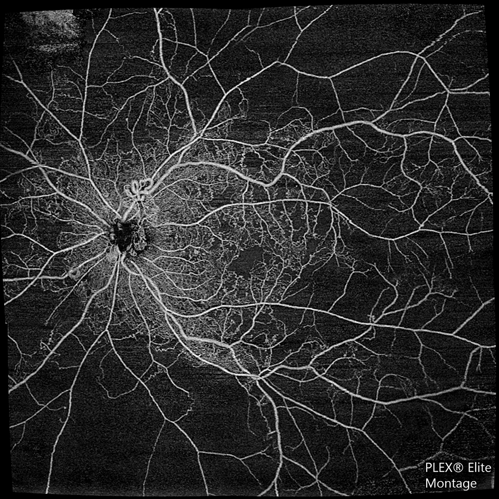

Ultra-widefield AngioPlex montage of Proliferative Diabetic Retinopathy, Superficial Retinal layer

Ultra-widefield AngioPlex montage of a case of IRFAN, Retina Depth Encoded layer
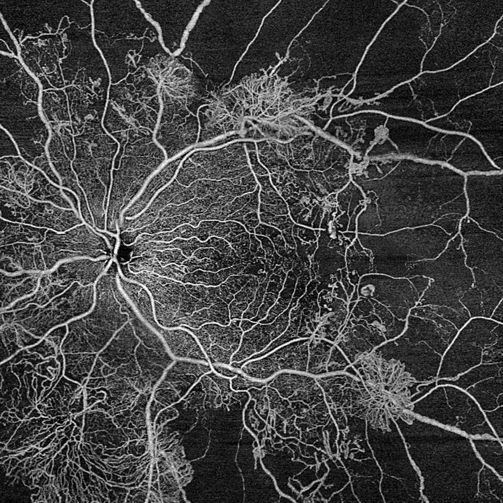


A Case of Proliferative Diabetic Retinopathy
This is a case of a young diabetic patient with severe proliferative diabetic retinopathy in both eyes. The new montage scan acquisition feature of the PLEX Elite 9000 is able to capture ultra-wide OCT angiography images that, in this case, helped the clinician clearly visualize the significant increase in the proliferation of new vessels in both eyes.
Drag the slider side-to-side to track disease progression in this DR patient over just four months. Slide to the right to see the patient’s first image. Slide to the left to see the second image, four months later.2
Videos


Drive the Discovery.
Join the Advanced Innovation and Research Network.Discovery starts with desire—the desire and drive to go where others have only imagined, to pave a path for those who will follow. At ZEISS, we're driving the discovery by investing in people, partnerships, and the emerging technologies that will share and define the future of patient care. With an entire global community of like-minded pioneers sharing our desire for discovery, our collaboration will continue to provide inspiration for years to come.

Where collaboration thrives, innovation emerges.
The Advanced Research and Innovation Network.
Downloads


Specifications
ZEISS PLEX Elite 9000 Swept-Source OCT Angiography-
-
MethodologyLine-scanning ophthalmoscope (LSO)Live fundus imageDuring alignment and during OCT scanOptical sourceSuper-luminescent diode (SLD), 750 nmField of view36º W x 30º HFrame Rate> 20 Hz
-
MethodologyCCD cameraResolution1280x1024
Get in touch with us!
Receive more information about the product and availability in your country!Related products


-
1
ZEISS PLEX Elite 9000 is CE marked, 510(k) cleared and is available for sale in selected countries and in the United States.
-
2
Images courtesy of Dr. Jean-François Korobelnik, University Hospital Pellegrin, Bordeaux, France.