You are on our international English website. This site features our entire product portfolio worldwide. The products featured may not be available in the US. If you are a citizen from the US, please visit your country website for local information and contacts.
You are on our international English website. This site features our entire product portfolio worldwide. The products featured may not be available in the US. If you are a citizen from the US, please visit your country website for local information and contacts.
Clinical challenges of Spinal Decompression Surgery
Spinal decompression surgery aims to relieve symptoms related to nerve compression in the affected regions of the spine. For nerve compressions in the cervical area, Anterior Cervical Discectomy and Fusion (ACDF) has emerged as a standard procedure for treating herniated discs. This technique involves the removal of damaged discs and bone spurs to relieve pressure on the spinal cord or nerve roots.1
One of the major challenges of ACDF surgery is the accurate identification of surrounding tissues. Factors such as visibility and the depth of the surgical field can hinder clear identification of the spinal cord and adjacent delicate structures,2 thereby increasing risk of spinal cord injury or nerve root damage.3
Depth+ for enhanced depth of field
Instant visual information on different anatomical layers is key to an efficient and safe procedure. With the new Depth+ mode of the ZEISS PENTERO 800 S, you can further increase depth of field for a higher resolution in the outer area of your current focal point.
Enabling you to see everything in your field of view sharply with just a click – whenever you need it.
Enhanced Resolution
Achieve sharp views of the entire region of interest with increased depth of field.
Surgical Precision
Confidently navigate deep anatomical channels while operating at higher magnification
Increased Efficiency
Minimize time spent on focus and zoom adjustments by effortlessly switching between Standard and Depth+ mode.
Comparison of Standard mode and Depth+ mode
Depth+ case example
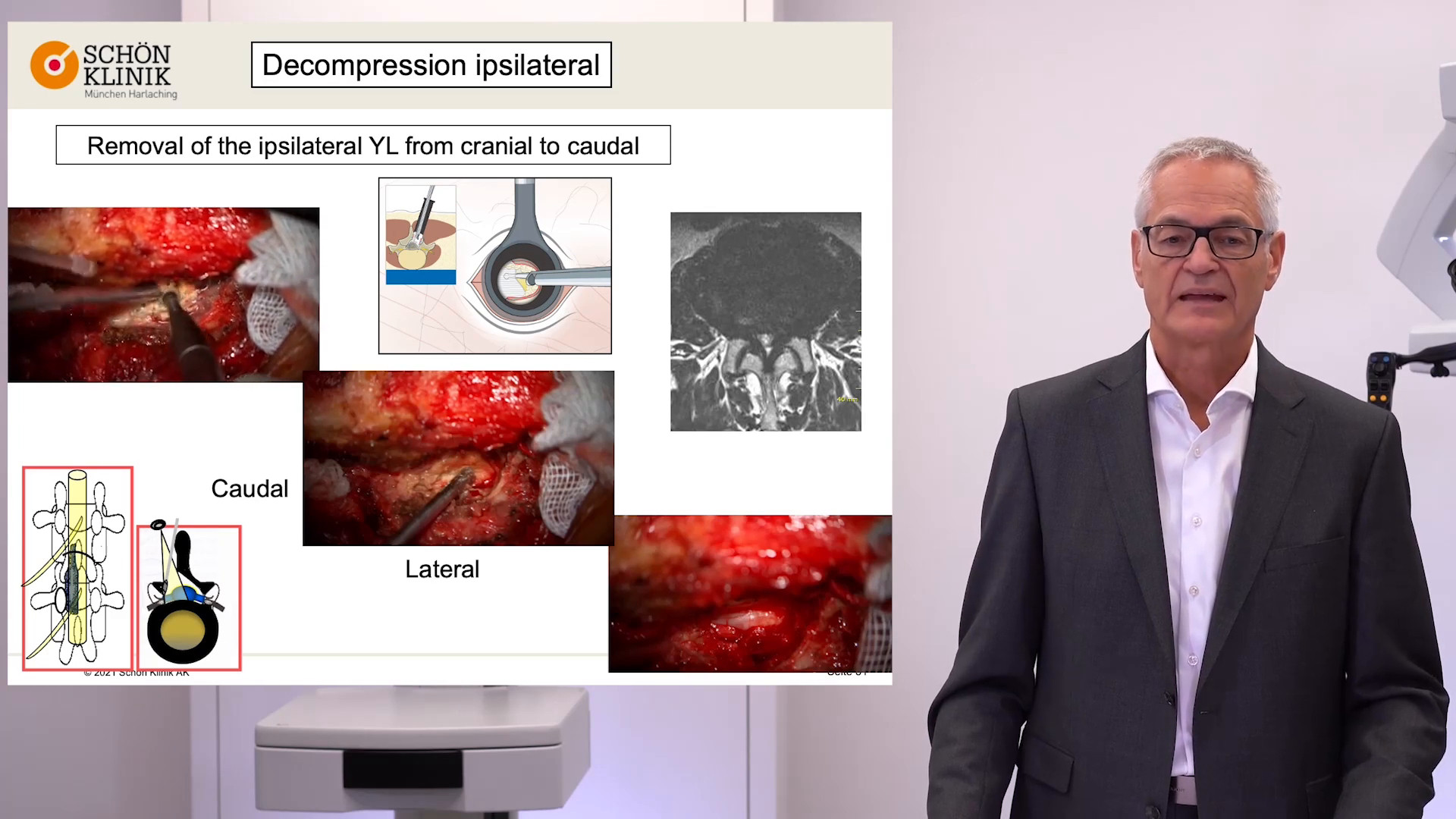
Anterior cervical decompression surgery
Compare the images of Standard mode and the Depth+ mode of the decompression surgery (anterior cervical spine surgery C3 - C4) and experience the difference in the depth of field. Thanks to the new Depth+ mode, the focus is on both the disc space and the anterior wall.
I realized while working on the cases that I was so accustomed to wanting more depth perception, that I was using less magnification. Normally, I use less magnification to be able to see more depth. However, with Depth+, I could magnify more without losing that depth perception.
First-hand experiences with Depth+
Third-party Content Blocked
The video player is blocked due to your cookie preferences. To change the settings and play the video, please click the button below and consent to use of "Functional" tracking technologies.
In this recorded webinar, Prof. Claudius Thomé shares his experiences with the ZEISS PENTERO 800 S concerning Depth+. In his talk, he presents first learnings from the use of the device as well as use cases.
Download Depth+ Infographic
Simply fill out the form below to receive the infographic.
Find out how the Depth+ mode integrated in the PENTERO 800 S from ZEISS will support your spinal procedures.
Advanced Visualization Systems from ZEISS
ZEISS PENTERO 800 S
Designed to suit your ambitious needs in spine surgery, neurosurgery as well as plastic & reconstructive surgery by offering Visual Certainty with extended optical capabilities, Seamless Performance with reinvented interactions, and Integrated Connectivity with leading digital solutions.
KINEVO® 900 S from ZEISS is the leading neurosurgical microscope platform. It leverages advanced robotic-assisted movement and positioning capabilities to enhance digital visualization.
During advanced surgical interventions and post-procedure workflows, TIVATO® 700 from ZEISS is your intelligent partner that enhances all-around performance when it is needed most.
You would like to explore further clinical experiences of your peers? Just register for MyZEISS to discover additional clinical articles and webinars in our Peer Insights area.
Ruetten, S. et al. Full-endoscopic anterior decompression versus conventional anterior decompression and fusion in cervical disc herniations. Int Orthop. 2008 Nov 18;33(6):1677–1682. doi: 10.1007/s00264-008-0684-y
2
Dong, Y. et al. The Clinical Efficacy of Anterior Cervical Discectomy and Fusion Under Three-Dimensional Microscopy. World Neurosurg 2024 Oct:190:309-310. doi: 10.1016/j.wneu.2024.07.182. Epub 2024 Aug 7. https://pubmed.ncbi.nlm.nih.gov/39097085/
3
Cheung, J. et al. Complications of Anterior and Posterior Cervical Spine Surgery. Asian Spine J. 2016 Apr 15;10(2):385–400. doi: 10.4184/asj.2016.10.2.385