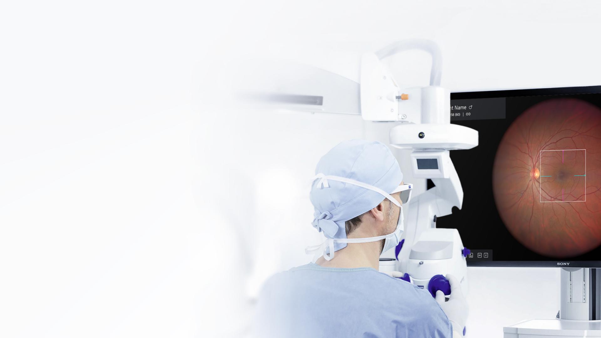

ZEISS ARTEVO 800
Experience more confidence during retina surgery.When it comes to retina surgery, the visualization capabilities of ZEISS ARTEVO 800 provide exceptional details even when using reduced light intensity. More than 75% of surgeons report a significant reduction in light intensity1, which can possibly result in lower phototoxicity.
The increased magnification and high-resolution OCT images allow you to more accurately identify and distinguish tissue structures of the retina – directly in your line of sight. That means you can make more informed decisions and perform with more confidence.


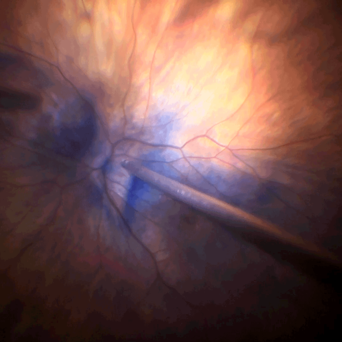
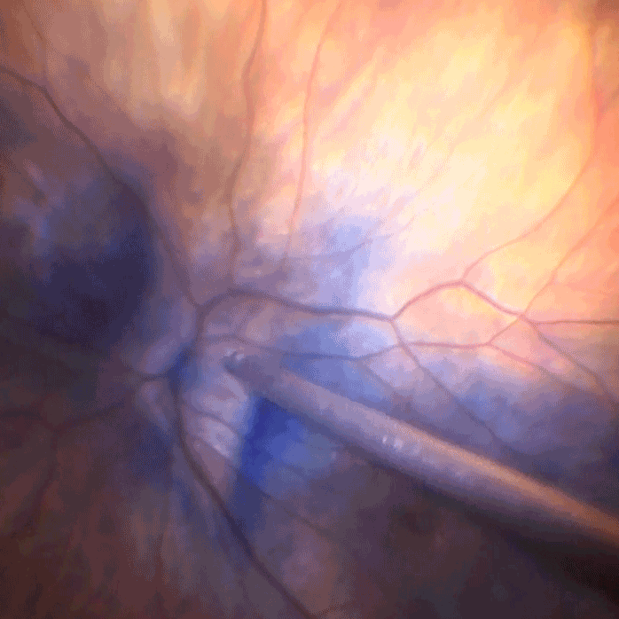
Reduce phototoxicity using lower light intensity.
For the possibility to decrease phototoxicity for your patients, you can reduce light intensity by up to 85%.1 This is made possible thanks to the enhanced visualization of DigitalOptics with two 4K cameras, providing natural colors and extraordinary resolution. No matter the light intensity you choose, you can still operate with efficient brightness without compromising the view.

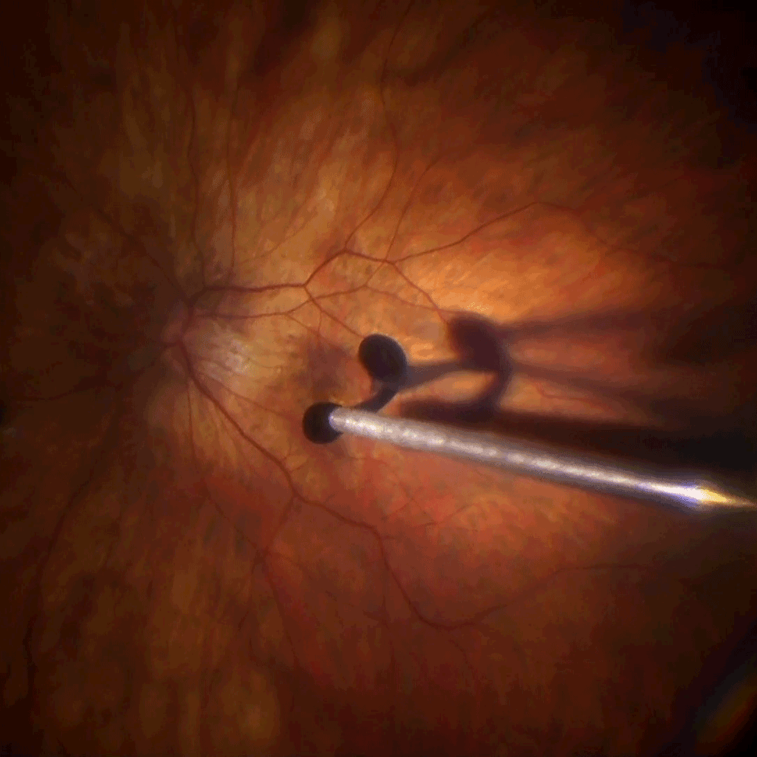


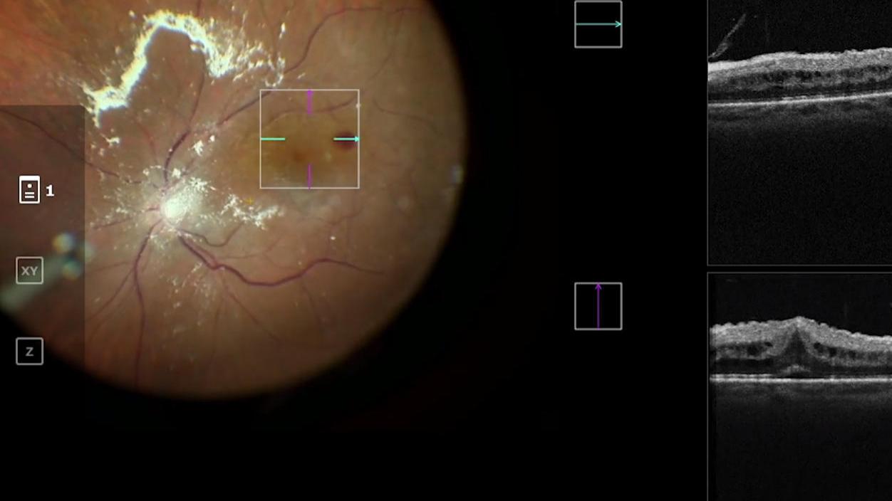

Make more informed decisions with integrated intraoperative OCT.
While OCT scans can reveal the retina’s distinctive layers and could help diagnose eye disorders, ZEISS ARTEVO 800 unleashes the full potential of intraoperative OCT images like never before. With 39% more image information1 and magnification at a new level, you can reveal significant insights, which allow strategy adjustments during surgery.
Intraoperative OCT can support during various retinal procedures. It can help to reveal epiretinal membrane, macular edema, posterior hyaloidal traction, and retinal detachment, for example.2
Get in touch with us!
Receive more information about the product and availability in your country!-
1
Data on file.
-
2
Ehlers, Justis P., Joseph F. Griffith, and Sunil K. Srivastava. “Intraoperative OCT during Vitreoretinal Surgery for Dense Vitreous Hemorrhage in the PIONEER Study.” Retina (Philadelphia, Pa.) 35.12 (2015): 2537.