You are on our international English website. This site features our entire product portfolio worldwide. The products featured may not be available in the US. If you are a citizen from the US, please visit your country website for local information and contacts.
You are on our international English website. This site features our entire product portfolio worldwide. The products featured may not be available in the US. If you are a citizen from the US, please visit your country website for local information and contacts.
Visualization methods in MISS: Microscope, endoscope or loupes?
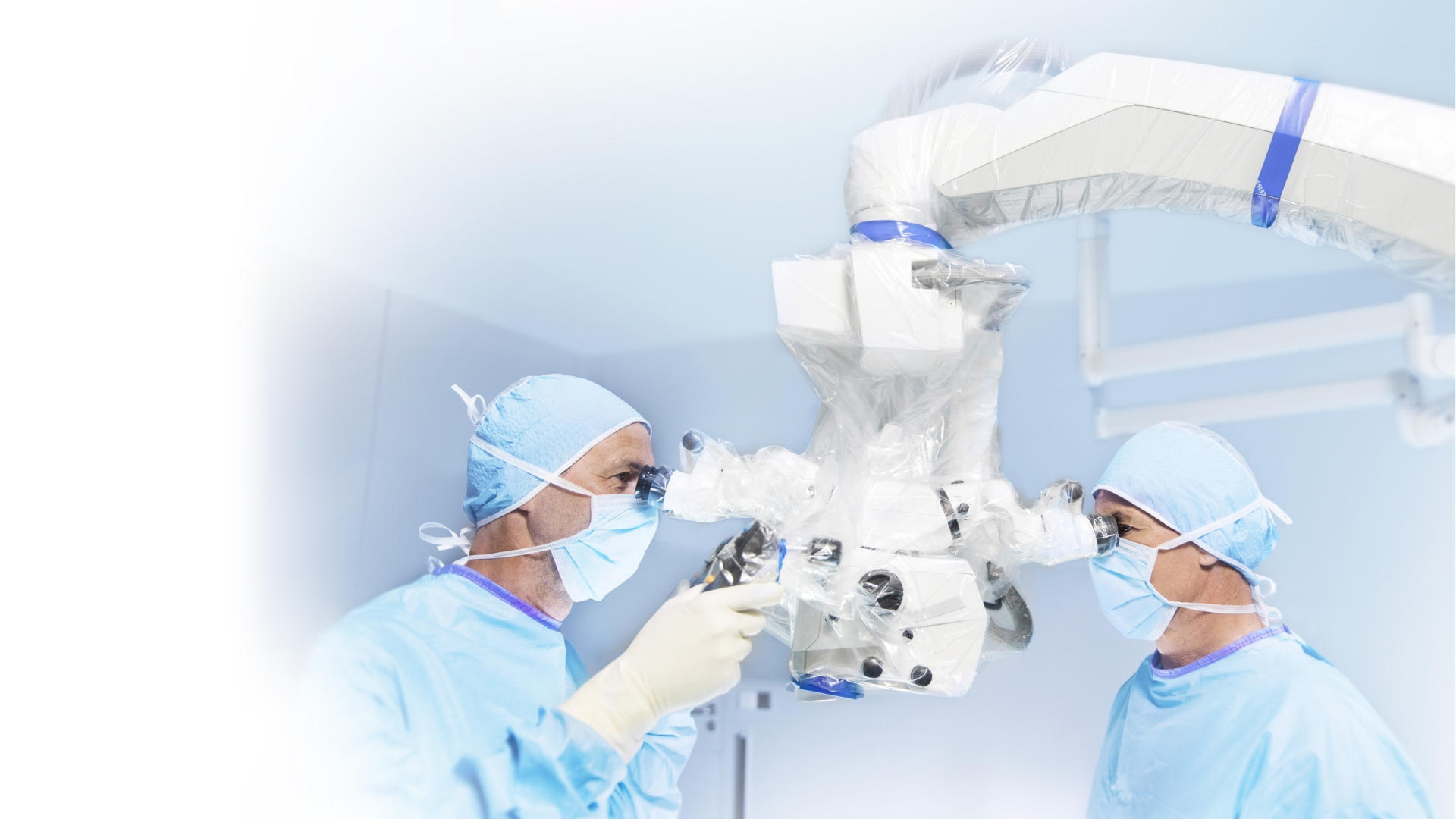
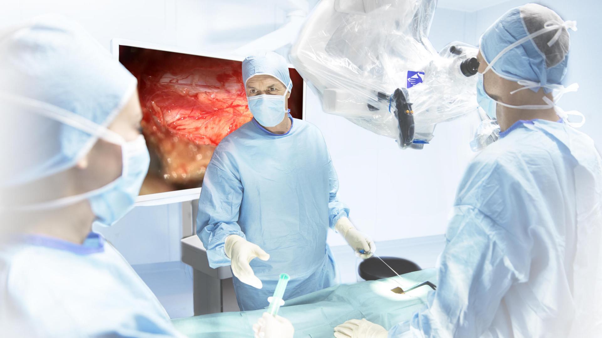
Visualization methods differ in terms of visualization quality, ergonomics provided to the spine surgeon as well as digital documentation properties. Compared to an endoscope, an Advanced Visualization System provides advanced magnification and illumination so that the surgeon can fully focus and treat the pathology without affecting surrounding healthy tissues.¹ The system also offers longer focal distance and better depth perception than an endoscope,¹ and doesn’t compete for space within the small surgical opening.
Increasingly, we are aware of the musculoskeletal impact of hours spent performing surgery. Over 60% of spine surgeons suffer from lower back pain, and 59% from neck pain.¹ That’s almost three times higher than the general population. Compared to loupes, an Advanced Visualization System allows the surgeon to stand upright, maintaining the neck and back posture and reducing strain.1 They are flexible and easy to maneuver2. Loupes offer no opportunity to share the surgeon’s view – a huge advantage of an Advanced Visualization System in the modern OR where a shared video screen allows any surgical team to follow the progress of the surgery, and students can observe and learn.¹
That‘s how the operative microscope has really changed my practice […]. I think it has a significant value not only from MIS adoption but then technical proficiency of the surgery as well as ergonomics. And I think as a result, fatigue factor […], live expectancy of a spine surgeon […] have improved significantly. And so it is certainly a work horse for me.
ZEISS SOLUTION
Advanced Visualization Systems for MISS from ZEISS
Microsurgery and the minimalized approach can be performed when using a visual aid and an optical system which allows the spine surgeon to work in a very narrow working corridor. For the choice of a visualization system different factors can be considered such as the surgeon’s needs and daily requirements, the surgeon’s specialty and the positioning in the operating theatre.
There are still challenges associated with MISS, e.g. proximity to spinal cord, working through a narrow channel with a small field of view, use of long instruments, the need for stability, and ergonomic issues if using loupes or no magnification. But choosing the right equipment supports the surgeon to improve results and patients’ outcomes.
A fully integrated Advanced Visualization System offers the following benefits:
Enhanced visualization – stereoscopic magnification and superior illumination2
Superior ergonomics – with flexibility and maneuverability so the surgeon can work in a comfortable upright position without straining the neck or lower back2
Digital documentation – so the surgeon can record, document and analyze
Wood M. The role of the surgical microscope in modern MISS. Becker’s Spine Review 2016. Available at: https://www.beckersspine.com/spine/item/32515-the-role-of-the-surgical-microscope-in-modern-mis-spine-drs-k-d-riew-michael-mayer-roger-haertl-mohd-hisam-muhamad-ariffin-share-their-experiences.html
2
Damodaran O et al. Microscope in modern spinal surgery: advantages, ergonomics and limitations. ANZ J Surg 2013;83:211–214