You are on our international English website. This site features our entire product portfolio worldwide. The products featured may not be available in the US. If you are a citizen from the US, please visit your country website for local information and contacts.
You are on our international English website. This site features our entire product portfolio worldwide. The products featured may not be available in the US. If you are a citizen from the US, please visit your country website for local information and contacts.
For many years, the operating microscope has established itself as a benchmark for intraoperative visualization in advanced microsurgery, delivering exceptional magnification and illumination of spinal anatomy and pathologies. Its capacity to offer detailed visualization has made it a vital element of modern surgical practices.1
However, despite these advantages, there are inherent challenges with relying solely on microscopic visualization:
Visibility constraints
The operating microscope's reliance on a straight line-of-sight can lead to missed critical information hidden behind tissue or corners, making it challenging to fully assess the surgical field and critical structures during procedures.2
Excessive dissection
The narrow field of view may require additional soft tissue and bony dissection to achieve adequate visualization, which can complicate the procedure and increase the risk of converting to open surgery due to inadequate access.3,4
Ergonomic strain
Adjusting the microscope to challenging angles for optimal viewing can lead to uncomfortable postures for the surgeon when working through the oculars only, causing physical discomfort and potentially affecting surgical performance and efficiency.5
These challenges are particularly pronounced in clinical applications such as minimally invasive spine surgery, where confined spaces make it difficult to position the microscope and instruments for optimal visualization.
ZEISS QEVO® provides efficient access to additional visual information
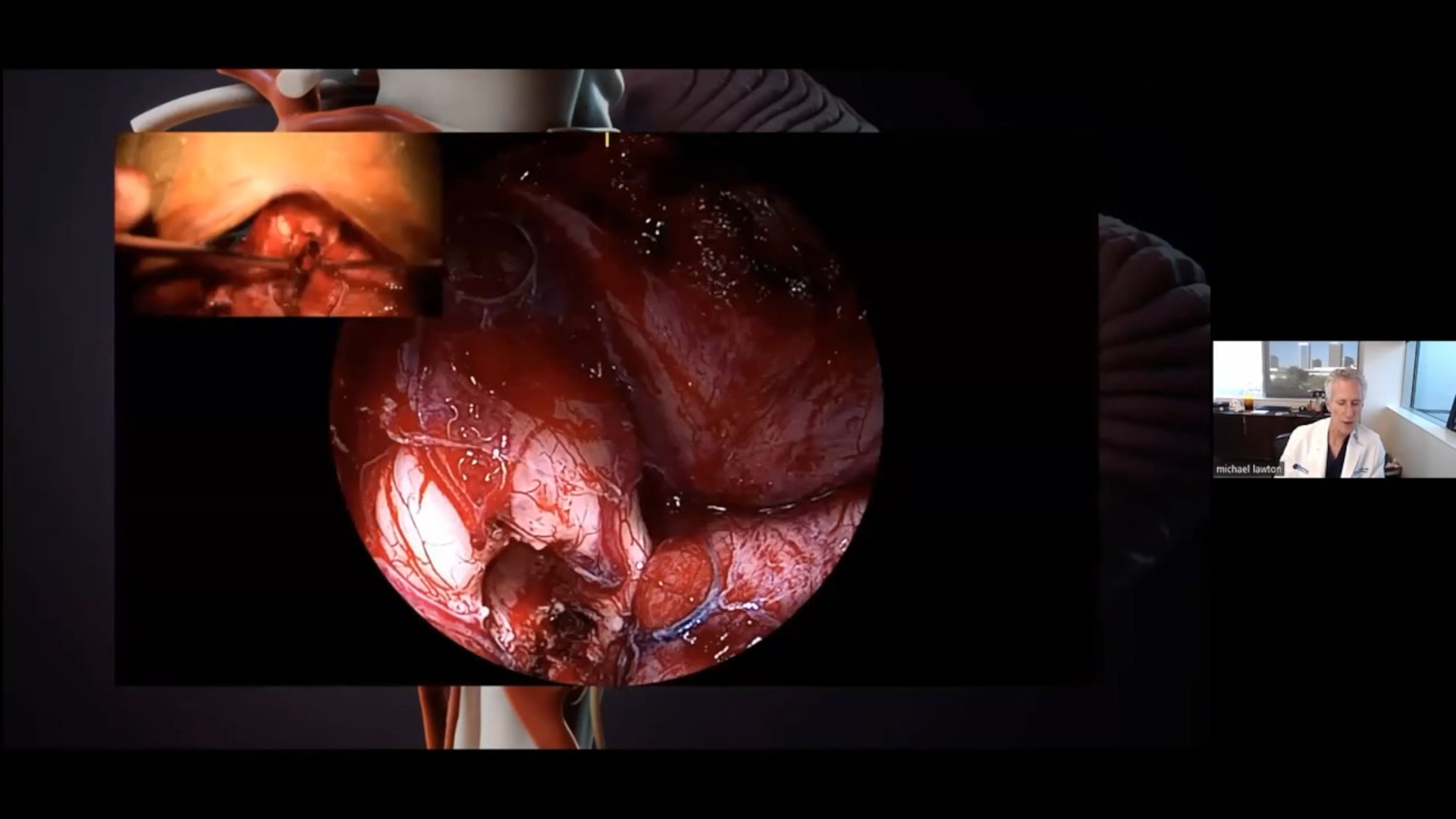
With the unique Micro-Inspection Tool QEVO from ZEISS, you can eliminate these blind spots in the microsurgical view and effectively look around corners. Whenever needed, plug it in to your ZEISS PENTERO 800 S to get an immediate endoscopic view on the anatomy without increasing the surgical footprint.
Providing efficient access to additional visual information – for greater certainty during your procedures.
Third-party Content Blocked
The video player is blocked due to your cookie preferences. To change the settings and play the video, please click the button below and consent to use of "Functional" tracking technologies.
ZEISS QEVO case example
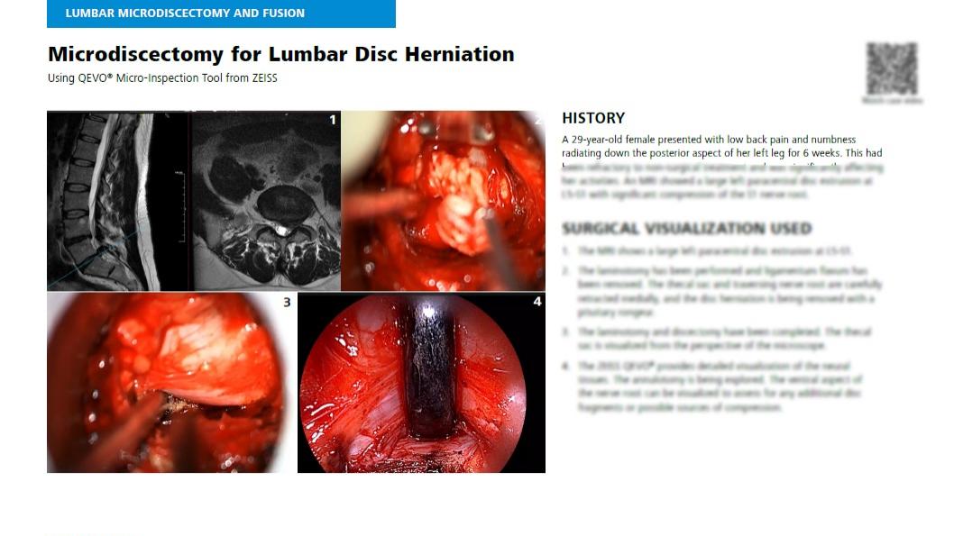
During a lumbar microdiscectomy, where herniated disc material is removed to relieve nerve root compression, the ZEISS QEVO offers detailed visualization of neural tissues, allowing surgeons to examine the ventral aspect of the nerve root for additional disc fragments or compression sources.
Interested in more details on this case?
Download the clinical case report
To receive more information on this case example (and 12 additional spinal cases in which the QEVO device proved especially useful) just fill out the form below and receive the full report – including clinical videos.
In the lumbar spine, when doing a decompression such as a microdiscectomy, I use the QEVO to completely visualize the nerve root and follow its path into the foramen to assess for any signs of compression or hidden disc fragments that I might not see with the microscope alone.
Simply fill out the form below to receive the eBook.
Discover 13 spinal cases where the QEVO device proved especially useful.
Advanced Visualization Systems from ZEISS
ZEISS PENTERO 800 S
Designed to suit your ambitious needs in spine surgery, neurosurgery as well as plastic & reconstructive surgery by offering Visual Certainty with extended optical capabilities, Seamless Performance with reinvented interactions, and Integrated Connectivity with leading digital solutions.
KINEVO® 900 S from ZEISS is the leading neurosurgical microscope platform. It leverages advanced robotic-assisted movement and positioning capabilities to enhance digital visualization.
During advanced surgical interventions and post-procedure workflows, TIVATO® 700 from ZEISS is your intelligent partner that enhances all-around performance when it is needed most.
You would like to explore further clinical experiences of your peers? Just register for MyZEISS to discover additional clinical articles and webinars in our Peer Insights area.
Moisi et al. Advancement of Surgical Visualization Methods: Comparison Study Between Traditional Microscopic Surgery and a Novel Robotic Optoelectronic Visualization Tool for Spinal Surgery. World Neurosurgery. 2017 Feb Pages 273-277. doi: https://doi.org/10.1016/j.wneu.2016.11.003
2
Nowak, S et al. Endoscope-Assisted Microsurgery for Posterior Fossa Skull Base Meningioma Surgery: Technique and Results. 2024 August, 27(2):137-147. doi: 1227/ons.0000000000001093
3
Zhaojie Chin BMed et al. Full-endoscopic versus microscopic spinal decompression for lumbar spinal stenosis: a systematic review & meta-analysis. The Spine Journal. 2024 June, Volume 24, Pages 1022-1033. doi: https://doi.org/10.1016/j.spinee.2023.12.009
4
Ridge et al. Heads-up Surgery: Endoscopes and Exoscopes for Otology and Neurotology in the Era of the COVID-19 Pandemic. Otolaryngol Clin North Am. 2020 Sep 29;54(1):11–23. doi: 1016/j.otc.2020.09.024
5
Alshabi et al. Exoscope Visualization, Navigation Guidance, and Robotic Precision in Spine Surgery. Journal of Minimally Invasive Spine Surgery and Technique 2025; 10(1): 22-33. doi: https://doi.org/10.21182/jmisst.2024.01508