

Assess
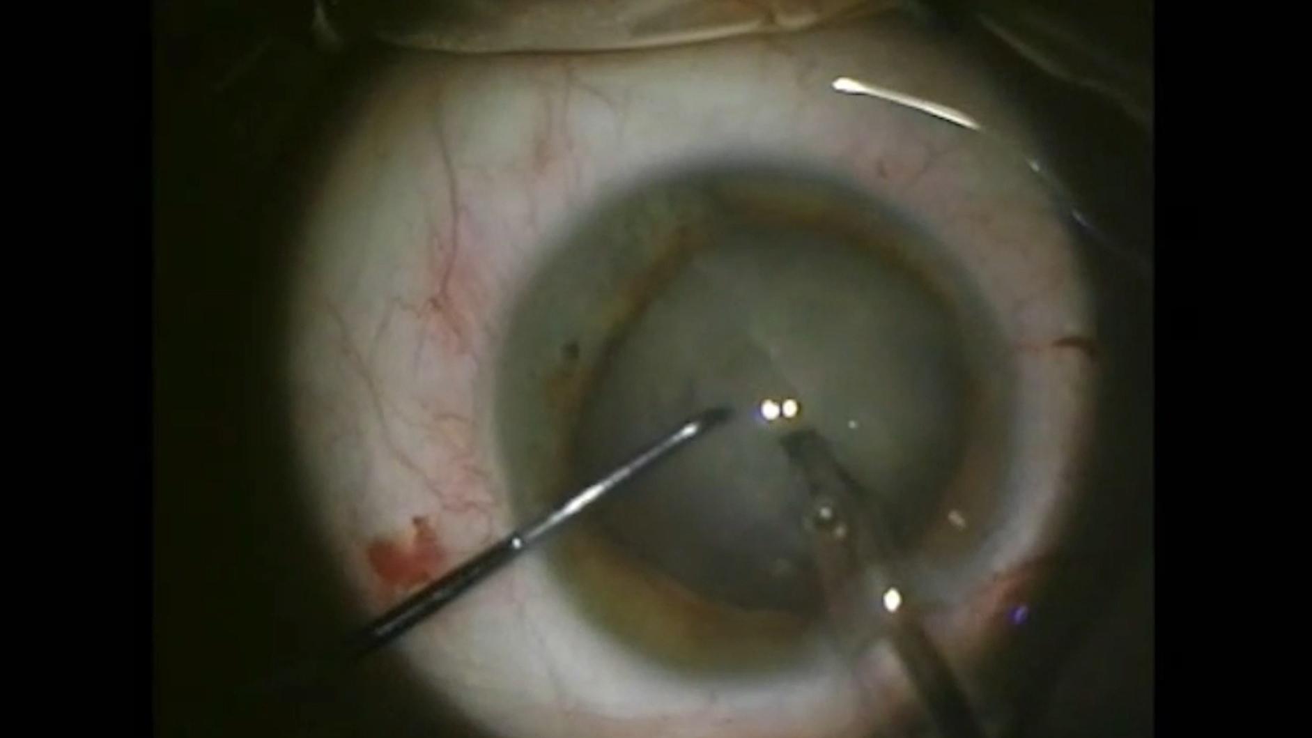
Dense cataract visualization and precise measurement

64-year-old female with a dense white intumescent cataract
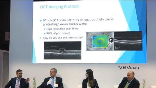
Screening out retinal pathologies in cataract surgery


ZEISS IOLMaster 700
The ZEISS IOLMaster 700 is designed to optimize workflow efficiency, even when handling dense cataracts, thanks to:
- Up to 99% cataract penetration rate, which reduces the need for ultrasound by 92%
- A measurement time of <45 seconds for both eyes7
- The unique Fixation Check to identify macular pathologies in your routine workflow8


ZEISS CIRRUS 6000
The ZEISS CIRRUS 6000 enables cataract practices to reveal hidden pathology and identify the right surgical candidates at the right time, leading to optimized workflow efficiency.
- Captures 100,000 A scans per second for a larger field of view of up to 12mm in a single scan
- Dense data acquisition protocols and oblique capture processes ensure quality diagnostic results
- Provides an objective status of the macula
- Helps set patient expectations around the final visual outcome of the surgery.


-
1
S.Garg, Surgeons meet challenges of removing rock-hard cataracts, Ocular Surgery News 2018, https://www.healio.com/news/ophthalmology/20181010/surgeons-meet-challenges-of-removing-rockhard-cataracts.
-
2
U. Devgan, Dense brunescent cataracts present surgical challenges, Ocular Surgery News 2011, https://www.healio.com/news/ophthalmology/20120331/dense-brunescent-cataracts-present-surgical-challenges.
-
3
A.Brissette, OCT Is Indispensable for Pre-op Cataract Evaluation, Opthalmology Management 2019, https://www.ophthalmologymanagement.com/newsletters/insights-into-integrated-diagnostic-imaging/may-2019.
-
4
Hirnschall N et al. Macular disease detection with a swept-source optical coherence tomography-based biometry device in patients scheduled for cataract surgery. J Cataract Refract Surg. 2016 Apr;42(4):530-6.
-
5
Hirnschall N et al. Enhanced Penetration for Axial Length Measurement of Eyes with Dense Cataracts Using Swept Source Optical Coherence Tomography: A Consecutive Observational Study. Ophthalmol Ther 2018;7:119-124.
-
6
R. Varsits, N. Hirnschall, B. Doeller, O. Findl; Increasing the number of successful axial eye length measurements using swept-source optical coherence tomography technology compared to conventional optical biometry; presented at ESCSR 2016.
-
7
Depending on eye condition and experience of the operator.
-
8
The ZEISS IOLMaster 700 is clearly not intended to be used for diagnostics. Findings need to be verified and pathologies diagnosed with a dedicated retina OCT.
-
9
M. Colvard, Phacoemulsification of the rock hard cataract, Eyeworld 2012, https://www.eyeworld.org/article-phacoemulsification-of-the-rock-hard.
-
10
http://corporate.ewreplay.org/?v=6122663037001.
-
11
R. J. Olson, MD, Salt Lake City, Review of Ophtalmology, PUBLISHED15 JANUARY 2005 Demystifying Dysphotopsia.