You are on our international English website. This site features our entire product portfolio worldwide. The products featured may not be available in the US. If you are a citizen from the US, please visit your country website for local information and contacts.
You are on our international English website. This site features our entire product portfolio worldwide. The products featured may not be available in the US. If you are a citizen from the US, please visit your country website for local information and contacts.
When cataract and glaucoma coexist, patients often seek care for cloudy vision from cataracts, while gradual vision loss from glaucoma may go unnoticed until advanced stages. Approximately 20% of cataract patients also have glaucoma or ocular hypertension.
Clinical solution
To determine if glaucoma affects a patient's vision improvement expectations and long-term vision related quality of life, it's essential to identify the glaucoma type, disease stage, and establish appropriate treatment plan.
ZEISS solution
ZEISS Glaucoma Workflow provides end-to-end solution for diagnosing monitoring, and treating glaucoma, including laser therapy and surgical options. The linked ZEISS Cataract Workflow improves outcomes for patients with both conditions.
Third-party Content Blocked
The video player is blocked due to your cookie preferences. To change the settings and play the video, please click the button below and consent to use of "Functional" tracking technologies.
“…and you can imagine the patients surprise, shock, disappointment and sadness when they have their cataract surgery and realize,
they don't regain as much vision as they want,
the vision they've lost is irreversible, and
they may have not had the most appropriate lens in their eye for their whole situation taken together.”
Dr. Matthew Schlenker
Glaucoma and Anterior Segment Specialist, Toronto Western Hospital, Canada
Step - Assess
Clinical Challenge: identify glaucoma types and risk factors
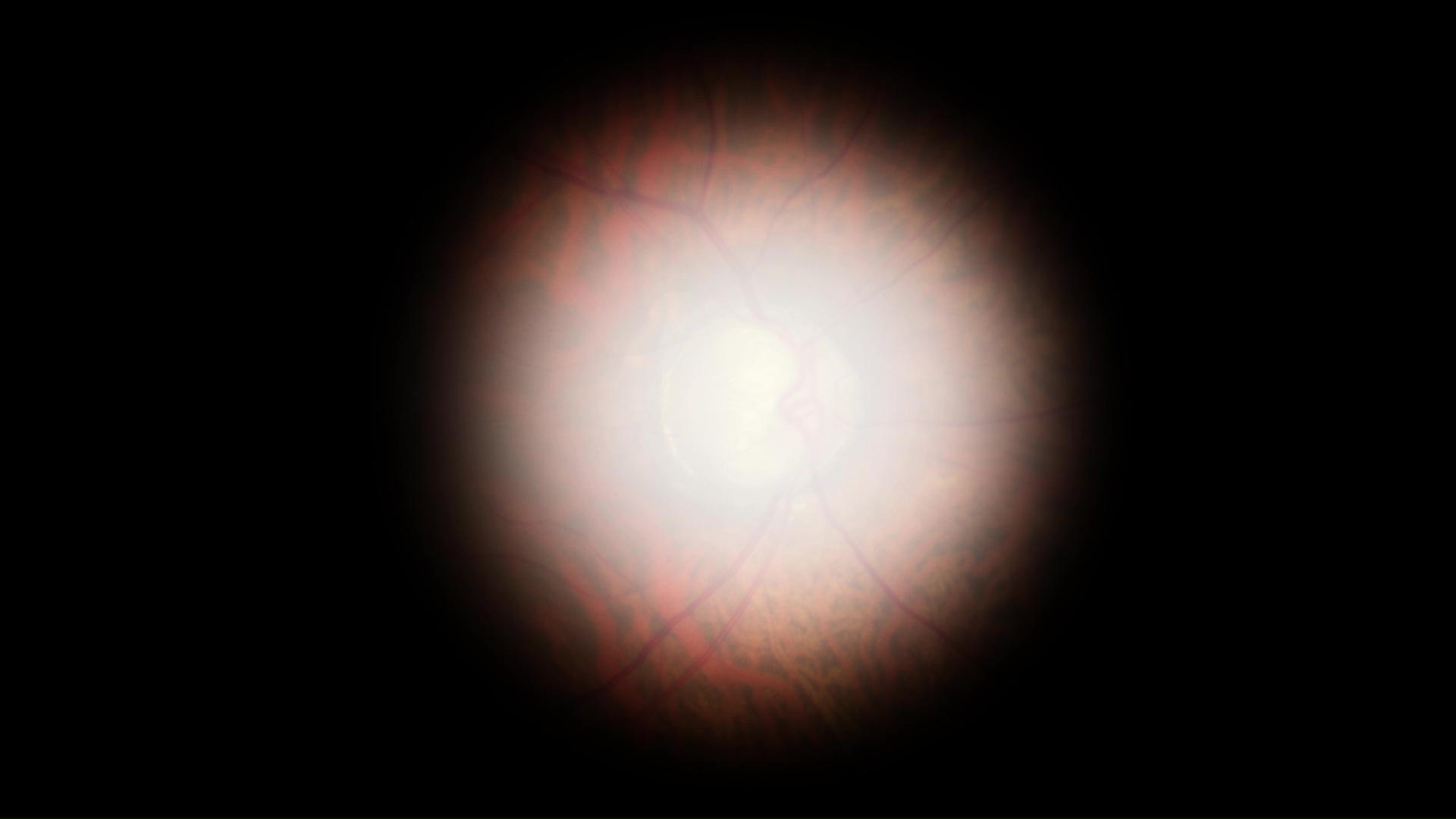
Slit-lamp ophthalmoscopy in cataract patients can detect glaucoma damage by evaluating the optic nerve and neuroretinal rim's size, color, and integrity.
Assessing anterior chamber aids in defining glaucoma type and recognizing “high-risk” eyes before cataract surgery.
Measuring intraocular pressure (IOP) before cataract procedure improves surgical care and supports long-term follow-up.
Solutions available from ZEISS:
“can be gained due to the TrueView Optics and VarioLight illumination of ZEISS SL 800.“ Ike Ahmed, MD, Prism Eye Institute, Canada
“With one hand, we can change the rheostat and lock the slit lamp as well. This is helpful to allow the other hand to do various maneuvers and to have one hand controlling the microscope itself.“ Ike Ahmed, MD, Prism Eye Institute, Canada
can be obtained using the Anterior Imaging Module of ZEISS CIRRUS 6000.
The ZEISS AT 030 tonometer mounts on the ZEISS SL 800, allowing other slit lamp extensions.
ZEISS’ biggest secret / surprise for anterior segment surgeons… ZEISS CIRRUS has full anterior imaging and quantification capability. So for folks who already have a ZEISS CIRRUS, you may already have the ability to perform epithelial thickness mapping (or upgrade your software to enable it).
Step - Plan
Clinical challenge: a plan for prompt and effective treatment
Glaucoma is a progressive disease that can negatively impact cataract surgery outcomes. Patient expectations, glaucoma type, and progression rate are key factors in selecting IOLs.
Combined CIRRUS and HFA analysis facilitates earlier and faster detection of glaucoma progression, supporting treatment before or during cataract surgery.
ZEISS Glaucoma Workplace integrates diverse data from ZEISS CIRRUS OCT, HFA perimetry, CLARUS fundus images, and IOP values for simplified visualization. In Progression Summary, the software highlights glaucoma changes across 5 test modes.
For evaluation before and after cataract surgery, ZEISS CIRRUS 6000 offers retina assessment, angle evaluation, and pachymetry in one device. ZEISS CIRRUS 6000 glaucoma analytics with GPA and an extended reference database, tailors glaucoma management to patient conditions.
The Guided Progression Analysis (GPA) of ZEISS HFA, the only FDA-approved method for assessing visual field loss progression, guides treatment decisions and estimates prognosis for vision-related quality of life.
What rates of HVF progression in glaucoma are clinically relevant?
”The fast progressors…those are the ones that are at high risk of going blind. If you have the MD change of -1.5 up to -2 decibels per year and -5% to -10% the equivalent in VFI, this is very fast, you should do something”.
Dr. Marta Pazos, Barcelona, Spain
FAST progresssors
MD change of -1.5 or -2 dB/year
VFI change of -5 to – 10%/year
CLINICALLY SIGNIFICANT rate of progression
Rate of progression that could lead to a high risk of future visual impairment for the patient.
FACTORS to be considered
Life expectancy.
Severity of disease.
Presence of known risk factors.
Everyday activities.
ZEISS solution: personalized glaucoma management
Third-party Content Blocked
The video player is blocked due to your cookie preferences. To change the settings and play the video, please click the button below and consent to use of "Functional" tracking technologies.
Observing differences in glaucoma progression is critical for individual vision health.
“It is important to evaluate the risk of disease progression and the likely rate of visual field loss. If we base our treatment decision on how each patient has done structurally and functionally, we are making a truly individualized risk assessment and measure prediction of vision related quality of life.”
Dr. Pilar Casas De Llera
Third-party Content Blocked
The video player is blocked due to your cookie preferences. To change the settings and play the video, please click the button below and consent to use of "Functional" tracking technologies.
Observing differences in glaucoma progression is critical for individual vision health.
"First, we want to have an accurate diagnosis of glaucoma. We need to stage the disease and then to assess the likelihood of progression. Depending on all these factors we will make the decision about what intraocular lens we should implant in each individual case."
Dr. Roberto Fernández Buenaga
Third-party Content Blocked
The video player is blocked due to your cookie preferences. To change the settings and play the video, please click the button below and consent to use of "Functional" tracking technologies.
Observing differences in glaucoma progression is critical for individual vision health.
“And we have these worrisome, Ganglion Cell complexes. So, the good news for this patient is we actually had a lot of data from the past and were able to utilize ZEISS Glaucoma Workplace to correlate the structure and function over time.“
Dr. Matthew Schlenker
Third-party Content Blocked
The video player is blocked due to your cookie preferences. To change the settings and play the video, please click the button below and consent to use of "Functional" tracking technologies.
Observing differences in glaucoma progression is critical for individual vision health.
“Before the surgery, if you use the visual field to explain to them what they can expect after the surgery, it will have a lot less trouble or complaint after the surgery with that patient.”
Dr. Manchima Makornwattana
Step - Treat
Clinical challenge (a): treat glaucoma prior to cataract surgery
For a glaucoma patient with cataract, treatment depends on glaucoma severity, visual field loss rate, cataract maturity, and overall health.
Pre-emptive SLT can reduce postoperative IOP spikes and medication dependency.
The ZEISS VISULAS® combi therapeutic laser, integrates photodisruption, photocoagulation, and selective photothermolysis without requiring lengthy adjustments or patient relocation, as the ZEISS VISULAS combi settings screen shows.
ZEISS SLT offers enhanced homogeneity, simplified titration, and better visibility of the trabecular meshwork. The laser energy selection is based on pigmentation that assists with reduction in unnecessary energy delivery.
Storing SLT treatment reports in ZEISS FORUM enhances clinical workflow efficiency. The VISULAS workstation can access ZEISS Glaucoma Workplace to review diagnostic data and monitor glaucoma treatment effects
Clinical challenge (b): combined glaucoma and cataract procedures
When the patient has coexistent glaucoma and cataract, a personalized approach should be taken.1
In cases with angle closure glaucoma, phacoemulsification can be effective for reducing IOP based on the EAGLE trial2
Phacoemulsification and glaucoma surgery can be combined for certain types of glaucoma patients to lower IOP and reduce the number of medications.
Intraoperative OCT in cataract and glaucoma procedures can help surgeons to observe surgical maneuvers in retina tissue and visualize devices during implant placement.
A first study showed a shorter total procedure time of QUATERA 700 compared to two competitive devices. Investigators graded intra-operative chamber stability was higher than for the two other systems.3
The ZEISS ARTEVO® 850 microscope integrates intraoperative OCT with visualization, adjustable for cataract, glaucoma or combined surgeries.
ZEISS solution: integration of cataract surgery and glaucoma management
Third-party Content Blocked
The video player is blocked due to your cookie preferences. To change the settings and play the video, please click the button below and consent to use of "Functional" tracking technologies.
Digital integration of diagnostics and treatment boosts workflow efficiency
…from patient prep and procedure to post-treatment assessment.
“As a glaucoma specialist, I can use SLT… I can use the possibility to perform laser peripheral iridotomy, iridoplasty, even gonio puncture after deep sclerotomy surgery. In addition to that, it has, the option to perform capsulotomy for secondary cataracts…and options for retinal treatment.”
Dr. Kaweh Mansouri
Third-party Content Blocked
The video player is blocked due to your cookie preferences. To change the settings and play the video, please click the button below and consent to use of "Functional" tracking technologies.
Digital integration of diagnostics and treatment boosts workflow efficiency
...from patient prep and procedure to post-treatment assessment.
“Integration of SLT with ZEISS FORUM means efficiency of our clinical workflow and the convenience of confirming the SLT results in front of the patient at the point of treatment.”
Dr. Pilar Casas De Llera
Third-party Content Blocked
The video player is blocked due to your cookie preferences. To change the settings and play the video, please click the button below and consent to use of "Functional" tracking technologies.
Digital integration of diagnostics and treatment boosts workflow efficiency
…from patient prep and procedure to post-treatment assessment.
“The ZEISS QUATERA has been almost transformative in how I do cataract surgery...One of the things that’s really nice about the ZEISS QUATERA unit, it allows us to remove cataracts in patients with shallow anterior chambers.”
Dr. Steven Vold
Third-party Content Blocked
The video player is blocked due to your cookie preferences. To change the settings and play the video, please click the button below and consent to use of "Functional" tracking technologies.
Digital integration of diagnostics and treatment boosts workflow efficiency
…from patient prep and procedure to post-treatment assessment.
“The ZEISS ARTEVO enhances the experience with its impressive 3-D viewing system and optics, supporting precise surgery.”
Dr. Matthew Schlenker
Step - Check
Clinical challenge: meet expectations for vision-related quality of life
Cataract surgery improves vision by removing a cloudy lens but doesn't cure glaucoma.
Careful evaluation, realistic expectations, meticulous surgery, and diligent follow-up can ensure patient satisfaction.
Patient data coming from multiple testing and treatment stations can be easily accessed via ZEISS FORUM®, a scalable and flexible ophthalmic data management platform that digitally connects all the workflow steps (Assess, Plan, Treat, Check) and links Glaucoma and Cataract workflows.
HFA3, CIRRUS and CLARUS devices deliver DICOM data into ZEISS FORUM for analysis in ZEISS Glaucoma Workplace. The SL-800 slit lamp and VISULAS laser from ZEISS transfer images and reports to ZEISS FORUM via Imaging Solution software. ZEISS solution from Dr. Pilar Casa De Llera perspective.
Multiple generations of ZEISS CIRRUS and HFA data can be used together in GPA, assuring full data continuity. ZEISS solution from Dr. Manchima Makornwattana perspective.
ZEISS Solution: continuous glaucoma care before and after intervention
Third-party Content Blocked
The video player is blocked due to your cookie preferences. To change the settings and play the video, please click the button below and consent to use of "Functional" tracking technologies.
Integrating ZEISS CIRRUS and HFA data in GPA
…to ensure ongoing glaucoma management and prevent vision loss.
“Glaucoma is a long-life disease and patients at first have no symptoms at all. So, if the patient doesn't know or isn't concerned about the problem at an early stage, they're going to have a problem which is permanent damage and irreversible.”
Dr. Manchima Makornwattana
Third-party Content Blocked
The video player is blocked due to your cookie preferences. To change the settings and play the video, please click the button below and consent to use of "Functional" tracking technologies.
Integrating ZEISS CIRRUS and HFA data in GPA
…to ensure ongoing glaucoma management and prevent vision loss.
“Let's say … we're able to stabilize this patient through either conservative measures or surgical measures. At that point, it's time to reset the baseline because we know that we have a better control of the patient's glaucoma. And we can do that right on ZEISS Glaucoma Workplace, right in front of the patient.“
Dr. Matthew Schlenker
Third-party Content Blocked
The video player is blocked due to your cookie preferences. To change the settings and play the video, please click the button below and consent to use of "Functional" tracking technologies.
Integrating ZEISS CIRRUS and HFA data in GPA
…to ensure ongoing glaucoma management and prevent vision loss.
“…We do believe that with the cataract surgery only…it will reopen the angle, make the eye pressure lower without doing anything more. And then we evaluate her again."
Dr. Manchima Makornwattana
Third-party Content Blocked
The video player is blocked due to your cookie preferences. To change the settings and play the video, please click the button below and consent to use of "Functional" tracking technologies.
Integrating ZEISS CIRRUS and HFA data in GPA
…to ensure ongoing glaucoma management and prevent vision loss.
“Laser treatments and minimal invasive glaucoma surgery will be the treatment of choice of these new generation of patients diagnosed early in the course of their disease. Regardless, glaucoma patients will need to be monitored for disease progression…Tools like ZEISS Glaucoma Workplace will help us in the management of such an increase of number of patients.”
Dr. Pilar Casas De Lliera
Watch your peers addressing the challenge of coexistent glaucoma and cataract
Managing cataract and glaucoma hand-in-hand
Challenges of angle closure glaucoma and the importance of the human touch.
Making surgical decisions for glaucoma comorbid cataract patients.
A bond of excellence: integrating glaucoma and cataract care.
Glaucoma journey from catching progression to surgical visualization.
Uncompromised care for cataract and glaucoma
Third-party Content Blocked
The video player is blocked due to your cookie preferences. To change the settings and play the video, please click the button below and consent to use of "Functional" tracking technologies.
Dr. Matthew Schlenker, Glaucoma and anterior segment surgeon
Dr. Marisa Sit, Comprehensive ophthalmologist
Toronto Western Hospital, Canada
Managing cataract and glaucoma hand-in-hand.
Third-party Content Blocked
The video player is blocked due to your cookie preferences. To change the settings and play the video, please click the button below and consent to use of "Functional" tracking technologies.
Dr. Manchima Makornwattana, Glaucoma and Cataract Specialist, Thammasat Univ. Hospital, Bangkok, Thailand
Challenges of angle closure glaucoma and the importance of the human touch.
Third-party Content Blocked
The video player is blocked due to your cookie preferences. To change the settings and play the video, please click the button below and consent to use of "Functional" tracking technologies.
Dr. Steven Vold, Glaucoma and Cataract Specialists, Vold Vision, Fayetteville, USA
Making surgical decisions for glaucoma comorbid cataract patients.
Third-party Content Blocked
The video player is blocked due to your cookie preferences. To change the settings and play the video, please click the button below and consent to use of "Functional" tracking technologies.
Dr. Pilar Casas de Llera and Dr. Roberto Fernández Buenaga
Fernández Casas Oftalmólogos, Torrelavega, Spain
A bond of excellence: integrating glaucoma and cataract care.
Third-party Content Blocked
The video player is blocked due to your cookie preferences. To change the settings and play the video, please click the button below and consent to use of "Functional" tracking technologies.
Dr. Matthew Schlenker, Glaucoma and Anterior Segment Specialist, Toronto Western Hospital, Canada
Glaucoma journey from catching progression to surgical visualization.
Third-party Content Blocked
The video player is blocked due to your cookie preferences. To change the settings and play the video, please click the button below and consent to use of "Functional" tracking technologies.
Prof. Dr. Koen Willekens, Visionair Oogzorg, Geel, Belgium
A guide on Surgical Innovations for Glaucoma, EGS, 2023
2
Azuara-Blanco A, Burr J, Ramsay C et al Effectiveness of early lens extraction for the treatment of primary angle-closure glaucoma (EAGLE): a randomised controlled trial. Lancet. 2016 Oct 1;388(10052):1389-1397. doi: 10.1016/S0140-6736(16)30956-4. PMID: 27707497.


 of ZEISS HFA")
 of ZEISS HFA")


