Image by Everaldo Coelho (Unsplash.com)

Focus on Microscopy 2023

2-5 April 2023 | Porto, Portugal

Learn about the latest microscopy innovations and connect with our experts at the Focus on Microscopy 2023. Explore the ZEISS Lattice Lightsheet 7 through hands-on technology demonstrations at our booth. Furthermore, you can experience our newest AI technology and scalable image analysis platform to get the best results out of your samples. Educational lectures and posters during the conference offer even more insights into the technology and applications of our imaging systems.

Demonstrations at our booth

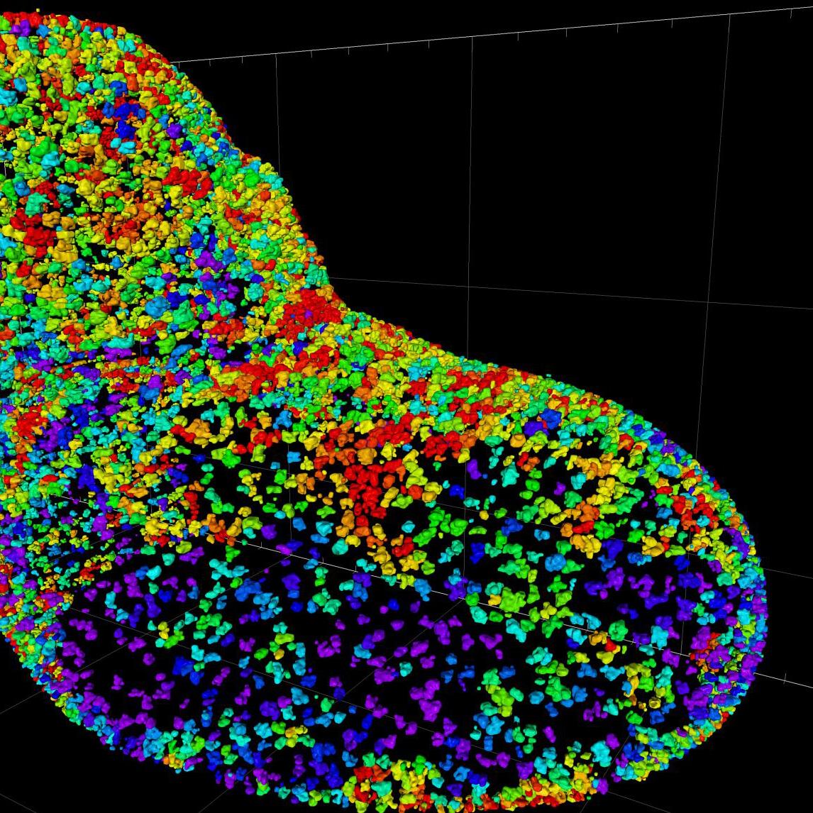
Experience live cell imaging at subcellular resolution with ZEISS Lattice Lightsheet 7

ZEISS Lattice Lightsheet 7 makes light sheet fluorescence microscopy available for live cell imaging at subcellular resolution – while also allowing you to use your standard sample carriers. With this automated, easy-to-use system, volumetric imaging of subcellular structures and dynamics over hours and days becomes available to everyone.
Discover the dynamics of life in unprecedented depth of detail by yourself during a hands-on demo session.

 Dataset provided by Anna Steyer and Yannick Schwab, EMBL
 Dataset provided by Anna Steyer and Yannick Schwab, EMBL
Dataset provided by Anna Steyer and Yannick Schwab, EMBL

AI technology for every scientist: The new integrated ZEISS image analysis platform

Image segmentation is one of the most crucial steps in the image analysis process. AI approaches can help with more robust and reproducible segmentation of even difficult samples, offloading a lot of tedious work from the scientist to the machine. We will present easy-to-use AI tools with which you can train your deep learning or machine learning algorithms that adapt to your research. No coding or expert knowledge is required and once the models are trained, they can be easily implemented in your image analysis workflows. The ZEISS image analysis platform offers ML and DL training and segmentation workflows that are supported locally and in the cloud, based on the proven APEER AI technology.

Scalable image analysis: Flexible data processing with Hub-based technology

Developments in organoid and spheroid research has increased the need of scalable tools for image analysis that can cope not only with 3D and 4D images but also with large size data sets. Hub-based data management provides a solution that can help any researcher to scale up their image analysis - regardless of dimensionality, image size, or complexity of the image analysis pipeline. The integration of Hub-based technology also includes AI-assisted approaches for image segmentation that can help any researcher to create robust image analysis workflows. Furthermore, the platform features collaboration and web-based access to data for peers, saving the researcher time copying data into multiple locations or devices.

Lectures and Posters

  • Speaker: Klaus Weisshart

    Time and Location: Monday, April 3, 14.20 – 14.40 pm, Infante Hall

    Abstract:
    Structured Illumination Microscopy (SIM) is a well-established method not only for optical sectioning but also super-resolution microscopy. In the classical SIM approach a 1-dimensional grid such as a line grating is used for structuring. Over the last years various 2-dimensional patterns have been employed leading among others to improved penetration depth.
    We will present how innovation on the encoding and decoding parts of structed illumination opens the possibility to distribute the photon budget according to the need of the experiment. With the example of ELYRA 7 we present data how reduced numbers of required raw images adapts the system perfectly to the special needs of capturing fast dynamics in living cells like for example organelle interactions. Moreover, sample-dependent resolution well below 100 nm can be achieved.

  • Speaker: Chris Power

    Time and Location: Tuesday, April 4, 16.10 – 16.30 pm, Archive Hall

    Abstract:
    Learn how to overcome the typical imaging and analysis challenges in neuroscience research, with the ZEISS LSM 9 series and arivis. LSM Plus and Airyscan joint Deconvolution (jDCV) can enable you to reliably gather higher quality data from your confocal imaging, enhancing resolution and signal to noise (SNR). Explore how the automatic end-to-end pipelines and AI-driven image analysis by arivis allow you to push the boundaries of your research. Automatic Neuron Tracing allows you to adapt the methods and parameters to your specific microscopy modalities and datasets, thus helping you to get the most out of your valuable imaging data from every sample.

  • Speaker: Kirstin Elgass

    Time and Location: Monday, April 3, 17:50-19:20 pm, Poster Hall

    Topic:
    Light sheet fluorescence microscopy has become an established method for fast and unsurpassed gentle imaging of living specimens. By adding lattice structures to the light sheet, ZEISS Lattice Lightsheet 7 makes this technique available for live cell imaging at subcellular resolution. The lattice light-sheet can be adapted to each experiment enabling visualization of previously unobserved events in the most challenging of samples, such as a single cell undergoing multiple mitotic waves, stem cell organoid development, malaria parasite invasion, and T-cell attacks.
    We will present technical details on the innovative approaches that turn Lattice Lightsheet 7 into an easy-to-use turn-key system leading to unique applications and research outcomes that demonstrate the potential for Lattice Lightsheet technology to drive new discoveries in biological and medical research.

Book your Demo here

Please note that you have to register yourself for the FOM conference via www.focusonmicroscopy.org upfront

Form is loading...

If you want to have more information on data processing at ZEISS please refer to our data privacy notice.

Meet our Experts

Meet our experts to learn how ZEISS can help to advance research, achieve insights, and minimize time-to-result with the latest in our portfolio of light and confocal microscopy solutions.

Chris Power

Product & Application Sales Specialist for LSM, Super resolution and Lightsheet systems in the EMEA region.

Delisa Garcia

Head of direct sales for EMEA and the Rest of World in charge of developing software business.

Sven Terclavers

Head of Product, Application & Sales Specialists EMEA-LA, leading an international team of microscopy specialists and business developers.  

Kirstin Elgass

Applications Development Engineer with Business Sector Life Sciences, Carl Zeiss in Jena, strategic product management, responsible for Special 3D systems.

Laura Machado

Portugal Sales Representative, focus on the sales of products and solutions of ZEISS Microscopy portfolio with active development of customers relationships.