


Join our free webinar on June 17th 2025
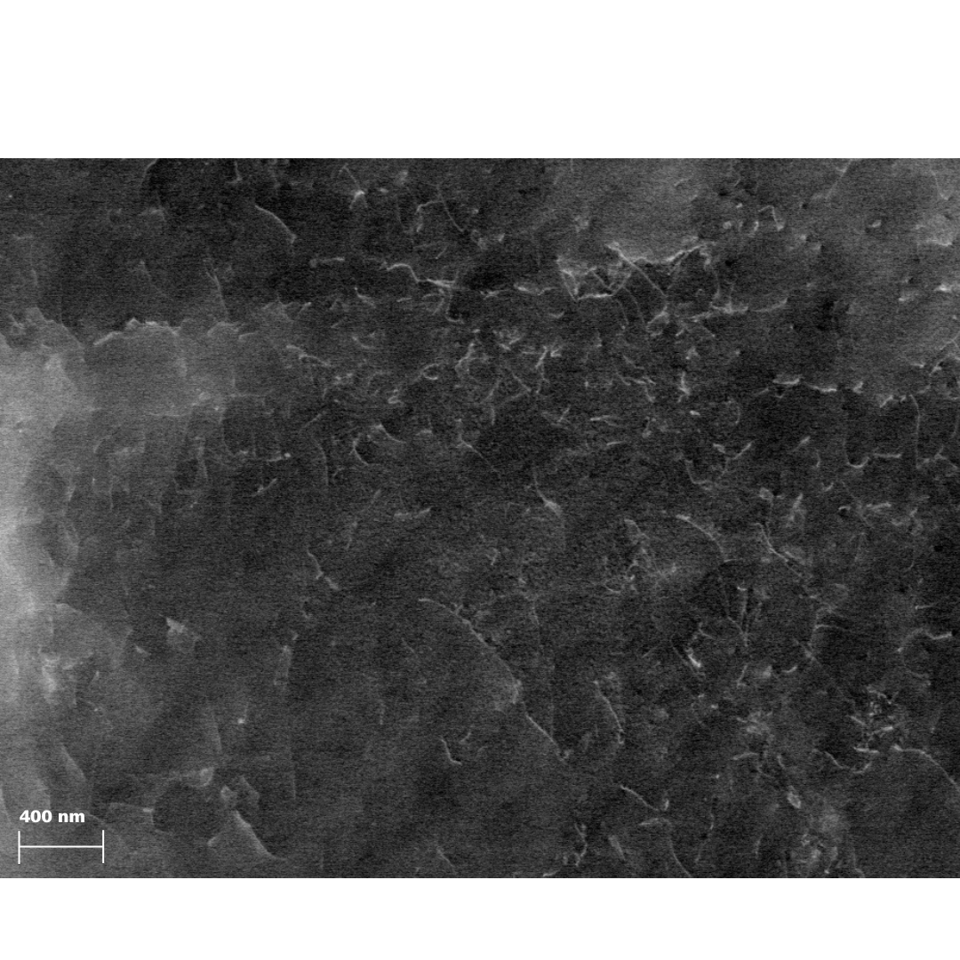
Controlled Electron Channeling Contrast Imaging (CECCI) In Scanning Electron MicroscopyDiscover a new approach to characterize dislocations in bulk samples using a scanning electron microscope (SEM) in this upcoming webinar. Explore how to visualize crystallographic defects in polycrystalline materials using controlled electron channeling contrast imaging (cECCI) in an SEM. Join us as we highlight the potential of cECCI and how it enables the observation of extended crystal lattice defects such as dislocations and stacking faults. It exploits the dependence of backscattered electron intensity on crystal orientation and atomic order.
Listen to a pioneer in materials science and study the basic principles of electron channeling contrast, the importance of determining the perfect imaging condition, and how any defect that disturbs the order of the lattice planes is made visible. Learn how to navigate the challenges of ECCI's low contrast intensity and master the necessary controlled workflow for optimal results.
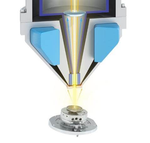
The method requires an SEM with optimal beam conditions and sophisticated crystallographic analysis software. The advantages over TEM (transmission EM) are that you are no longer limited to thin films as you can now observe bulk samples. This allows you to benefit from simplified sample preparation, facilitated in-situ experiments, and access to true sample representivity.
Don't miss this opportunity to enhance your understanding of the basic principles of cECCI utilizing a ZEISS field emission SEM with Gemini electron optics and TOCA (Tools for Orientation Determination and Crystallographic Analysis) software.