Live Webinar

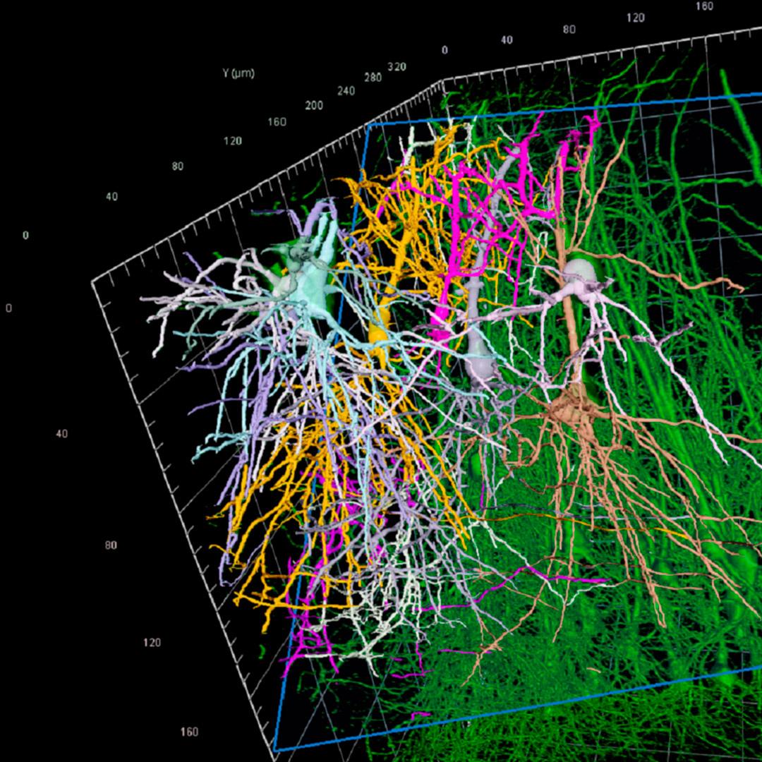
Neuroscience Research Without Compromises

Increase your productivity with faster results from better images

4 PM CEST / 10 AM EDT | Webinar
Starting soon
Ongoing
Event has Ended
  • 00 years
  • 00 months
  • 00 days
  • 00 hours
  • 00 minutes
  • 00 seconds

Abstract

As a neuroscience researcher, you are often faced with imaging quality issues, large data sets, long processing times, and limited computing power. At ZEISS Microscopy we have developed equipment and software tools that help you overcome these hindrances, and power up your workflows. LSM Plus and Airyscan joint deconvolution (jDCV) enable you to reliably gather higher quality data from your confocal imaging, enhancing resolution and signal to noise. Following the acquisition, automated end-to-end pipelines and AI-driven image analysis allow you to conduct experiments that traditional methods do not enable, and to do so on a whole new scale.

In this webinar, our microscopy and image analysis experts, Dr. Kalliopi Arkoudi and Dr. Delisa Garcia, will discuss the typical challenges researchers in neuroscience face. Image acquisition and analysis tips will be shared and demonstrated. Our new neuro tracing algorithms created to handle big data processing and overcome poor SNR will be presented.

Join us on October 20, 2022, at 4 PM (CEST) / 10 AM (EDT).


Key Learnings

In this webinar, you will learn how to enhance your image quality, and how to efficiently analyze data without compromises by:

  • Understanding imaging challenges such as Signal-to-Noise Ratio (SNR), resolution, and speed
  • Learning how LSM Plus can help you enhance SNR at high acquisition speed and low laser power
  • Exploring how Airyscan jDCV pushes resolution further while keeping sensitivity high
  • Processing images with poor SNR and big data using our new neuron tracing algorithms
  • Setting up automated pipelines for neuron analysis using arivis Vision4D, our image-type agnostic platform for advanced visualization and image analysis
Speaker Dr. Delisa Garcia Head of Sales (EMEA and ROW) – arivis, a ZEISS company

Dr. Delisa Garcia began using microscopy techniques such as confocal, multiphoton, second harmonics, FLIM, and FRET to study muscle contraction during her Ph.D. and postdoc at Imperial College London. In 2009, she left academia to advance her career in industry and has over ten years of experience in image handling and analysis in a wide variety of fields. She joined the arivis team three years ago as Head of Sales (EMEA and ROW), opening the doors to new approaches for image analysis using VR and AI.

Speaker Dr. Kalliopi Arkoudi Applications Development Engineer – ZEISS Microscopy

Dr. Kalliopi Arkoudi has a Master’s degree in Neuroscience from King’s College London (KCL), where she worked for three years on the characterization of interneurons in the zebrafish brain using confocal microscopy. She completed her Ph.D. in Inflammation and Regenerative Medicine in KCL, where she used various microscopy techniques including multiphoton imaging, FLIM, and FRET to study the importance of inflammatory pathways in the response of macrophages to injury. In 2020 Dr. Kalliopi Arkoudi joined ZEISS Microscopy as an Applications Development Engineer, where she is using her expertise in the development of high-end imaging applications for confocal microscopy.

Register for the Webinar

October 20, 2022 | 4 PM CEST / 10 AM EDT

Form is loading...

For more information on data processing at ZEISS, please refer to our data privacy notice.