

Characterize Biomaterials & Bio-Inspired Materials in 3D & 4D
Three studies illustrate how microscopy advances biomaterials science and engineeringDiscover three studies that illustrate how microscopy techniques support analysis of biomaterials and bio-inspired materials: From studying cartilage-bone interfaces in 4D, to the investigation of polymer fibers with spider-silk-like properties to the 3D characterization of nanofibrous scaffolds for tissue engineering. Scroll down and explore the use cases.
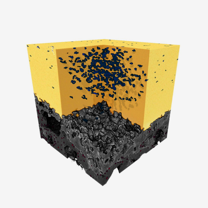

4D Investigation of Microdamage Initiation & Progression in Cartilage-Bone Interfaces
With X-ray tomographyIn order to understand failure mechanisms in biological tissues, X-ray Computed Tomography (XCT) enables researchers to track microdamage initiation and progression. In this study, Gianluca Tozzi from the school of Mechanical and Design Engineering at the University of Portsmouth investigates the mechanical performance & interplay of a cartilage-bone interface.
The goal: Contribute to the understanding of the etiology and pathogenesis of osteoarthritis, affecting millions of people worldwide.
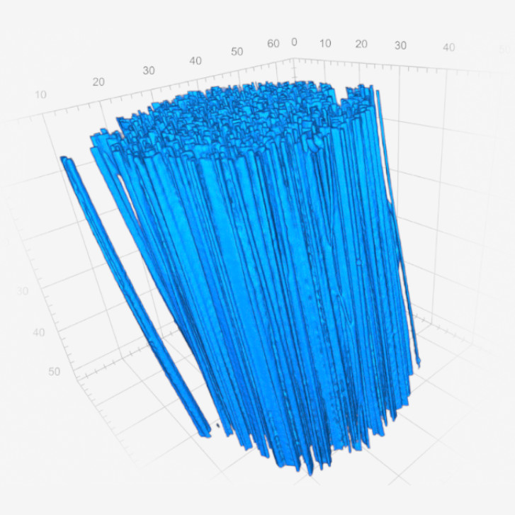

Characterization of Polymer Fibers with Dragline Spider Silk Properties
With X-ray tomography and FE-SEMScientists are trying to find a way to produce synthetic materials with a strength and toughness comparable to that of spider silk. To support the design of such a novel polymer fiber, researchers at the University of Halle-Wittenberg and the University of Bayreuth conducted microscopic characterization of a novel polymer fiber using SEM and XRM.
The goal: Reaching a better understanding of the link between microstructure, process and property.
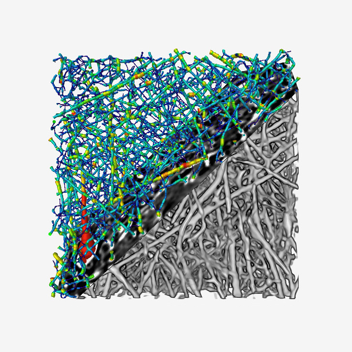

3D Microstructure Analysis of Nanofibrous Scaffolds for Tissue Engineering
With X-ray microscopyIn this study, researchers from the Divison for Microstructure based Materials Design (mikroMD) at University of Halle-Wittenberg and Fraunhofer IMWS investigated gelatin nanofibers, using X-ray microsopy to analyze their porosity, pore size & morphology.
The goal: Gain beneficial insights for the design and fabrication of novel fibrous materials for tissue engineering.