Innovators in Energy Research Lead the Way Towards Sustainability
Explore Microscopy Solutions for Energy Materials Research
Third-party Content Blocked
The video player is blocked due to your cookie preferences. To change the settings and play the video, please click the button below and consent to use of "Functional" tracking technologies.
In this interview, Prof. Joachim Mayer, RWTH Aachen, talks about materials as technology enablers and what we can expect to see in energy consumption and storage over the coming years.
Imagine the impact if we would find new ways for more sustainable energy technologies. Research and development of efficient devices such as batteries, solar cells, and fuel cells is crucial here, however, making this transition presents challenges.
Discover how innovators use advanced microscopy to reveal the connections between device and material microstructures and ultimate performance in these next generation technologies and point the way towards new breakthroughs in Energy Materials research.
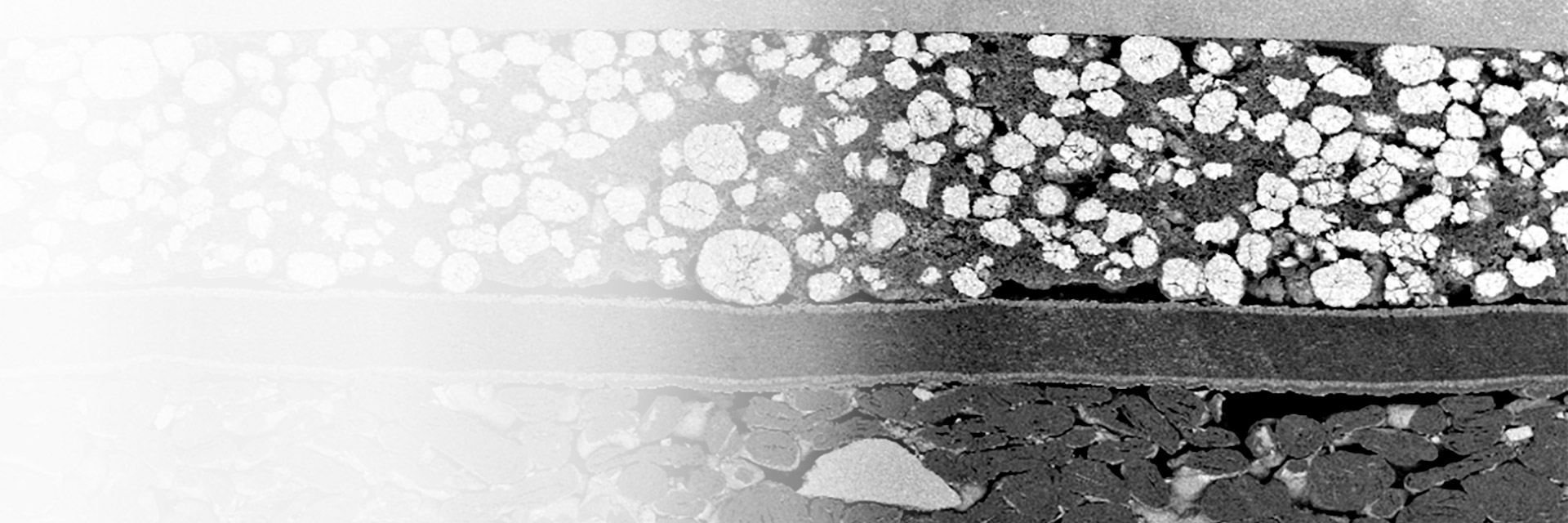
Multi-modal characterization of a solid-oxide fuel cell (SOFC) stack
The image here shows multi-modal characterization of a solid-oxide fuel cell (SOFC) stack with unwanted cracks. SOFC electrolytes should be pore-free while the electrodes need porosity so gas can reach the catalyst. Fuel cells supplied electricity on the 1960s Apollo missions but even today improvements are needed. As SOFCs operate in severe conditions at elevated temperatures, microcracking due to thermal expansion can lead to early failure. Correlative microscopy offers a way out here because several analytical methods can be applied together in one work step.
For further reading: Wolz, BC, Jaremenko, C, Vollnhals, F, Kling, L, Wrege, J, Christiansen, S. X-ray microscopy and automatic detection of defects in through silicon vias in three-dimensional integrated circuits. Engineering Reports e12520, (2022): https://doi.org/10.1002/eng2.12520
Prof. Silke Christiansen
Fraunhofer Institute for Ceramic Technologies and Systems IKTS
Energy systems like solar cells, fuel cells, and batteries contain many materials with varying properties, scales, and arrangements. Our goal is to control and optimize material and interface properties from macro to atomic scale with device manufacturers who optimize and control device performance. In our lab we established a comprehensive energy materials workflow which includes sample preparation, inert transfer, cryogenic environments, and full microscopic, spectroscopic, and bulk characterization using electrons, ions, x-rays, light, and probes in 2D and 3D. All this is connected so data can be analyzed correlatively and quantitatively using statistical tools and image recognition software powered by machine learning to help us create a digital twin and intelligently drive materials and device optimization.
Our goal is to optimize energy materials properties from the macro to the atomic scale. We established a comprehensive workflow using electrons, ions, X-rays, light and probes in 2D and 3D. All is connected so data can be analyzed using software powered by machine learning to help us create a digital twin and intelligently drive materials optimization.
The image here is from a recent paper on imaging and modelling of advanced Li-ion batteries. Using high resolution X-ray, ion-beam, and electron microscopy techniques, we gain insight into the huge complexity of these devices. There is a critical relationship between material morphology and device performance – for example in the trade-off between energy and power density in batteries. With an improved understanding of the fundamental relationship between device performance and microstructure, we are increasingly equipped to tackle these complex challenges.
Image courtesy of: Lu, X., Bertei, A., Finegan, D.P. et al. 3D microstructure design of lithium-ion battery electrodes assisted by X-ray nano-computed tomography and modelling. Nat Commun 11, 2079 (2020). Provided by Springer Nature. https://doi.org/10.1038/s41467-020-15811-x, licensed under CC BY 4.0: https://creativecommons.org/licenses/by/4.0/
Prof. Paul Shearing
University College London (UCL)
Microscopy techniques are central to our research – we have spent the past decade building a library of data on materials, electrodes, and electrochemical devices with our microscopes running around the clock. This is a real exemplar of a multi-scale challenge and provides exciting opportunities for correlative microscopy. With increasingly sophisticated tools for in situ and operando microscopy, we believe that we can condense the time it takes from discovering exciting new materials, to their deployment in the real world.
In The Electrochemical Innovation Lab at UCL, we help drive towards a Net Zero carbon future by studying electrochemical power systems including batteries, fuel cells, and super-capacitors in order to improve performance, durability, and safety, and lower cost.
The image above shows a silicon-based anode material imaged using one such workflow correlating X-ray microscopy and focused ion-beam scanning electron microscopy in a connected software environment. Green Areas indicate the correlated volume of interests (VOIs) and arrows indicate the workflow steps. To understand the morphology of Si-based anodes we need high contrast nm-scale resolution and a field of view at the µm-scale. Correlative imaging approaches like this unveil the underlying structural and chemical evolution in 3D and are highly beneficial for battery research.
The image above shows a silicon-based anode material imaged using one such workflow correlating X-ray microscopy and focused ion-beam scanning electron microscopy in a connected software environment. Green Areas indicate the correlated volume of interests (VOIs) and arrows indicate the workflow steps. To understand the morphology of Si-based anodes we need high contrast nm-scale resolution and a field of view at the µm-scale. Correlative imaging approaches like this unveil the underlying structural and chemical evolution in 3D and are highly beneficial for battery research.
In batteries, for instance, the electrochemical properties and morphology of the materials directly impact performance. Therefore, a deep understanding of structure-property relationships across length scales is key to improving the devices and materials within. Silicon-based anode materials hold promise for next-generation Li-ion batteries due to the high theoretical capacity, but challenges remain around capacity fade and cell life expectancy. Proper material engineering is essential here and an accurate representation of the complex multi-phase microstructures helps guide materials synthesis and processing efforts.
To do this we develop and apply advanced imaging and analysis workflows leveraging artificial intelligence to characterize the structural and chemical properties across cell- and microstructure-levels from mm to nm in 2D as well as 3D.
In my group we focus on image-based material characterization and analysis in energy storage and microelectronics. To do this we develop and apply advanced imaging and analysis workflows leveraging artificial intelligence to characterize the structural and chemical properties across the cell- and microstructure-levels from mm to nm in 2D as well as 3D.
Learn More on How Microscopy Can Be Applied in Energy Materials Research
In this 42-page WILEY ebook, get an overview of:
Microscopy techniques & their benefits for energy materials research
Microscopy-enabled accomplishments in energy materials research (11 short versions of selected peer-reviewed articles from various WILEY journals)
Current challenges & future developments in microscopy applications for energy materials (Interview with Professor Paul Shearing from the University College London)

 stack")
 stack")


