

Advanced Image Analysis Made Easy with ZEISS arivis
High-Performance Insights from 2D to 4D for Research and DiscoveryDiscover Image Analysis Possibilities with ZEISS
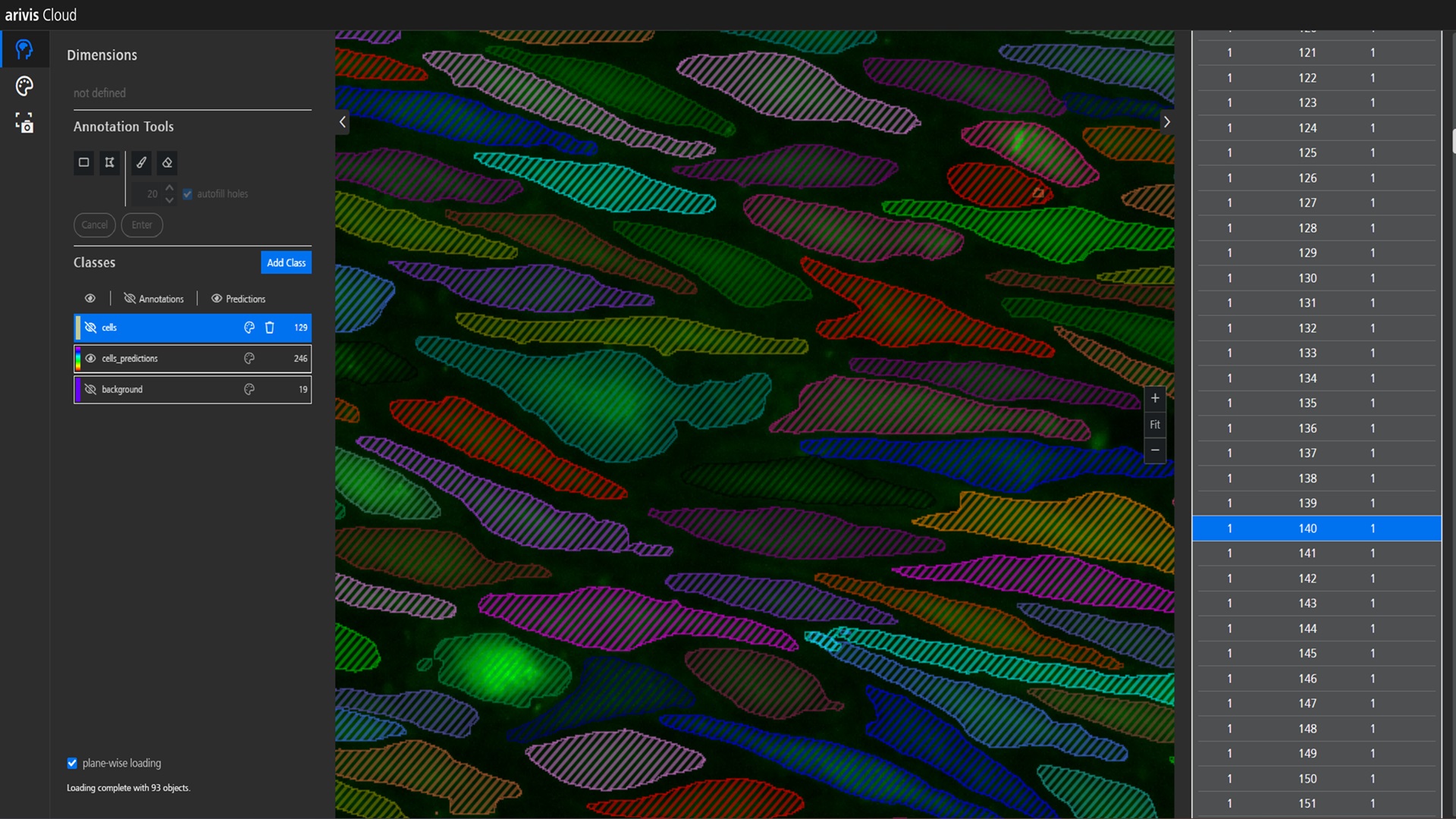
AI for Image Analysis: Power Made Simple
Train, customize, and deploy Deep Learning models - no coding required
Unlock the potential of AI with ZEISS arivis. It offers both cloud infrastructure and local options, coupled with an intuitive AI model training interface, making customized Deep Learning accessible to all. Its human-centered model training simplifies the process, requiring minimal image annotations to initiate training. Once created, models deliver standardized, reproducible assay results that eliminate variability between experiments and researchers.
- Accessible custom Deep Learning
- Instance (object-based) & semantic (pixel-based) segmentation
- Support for open-source models, such as Cellpose
- Support for diverse microscopy technologies and image formats
- DeepD3 for automated spine tracing in neuroscience research
- Improved standardization and reproducibility in 3D assays
- Seamless integration and automation
- No need to code
With ZEISS arivis, we built the image analysis platform we always wished for – one that empowers researchers to customize, automate, and scale their workflows with AI.

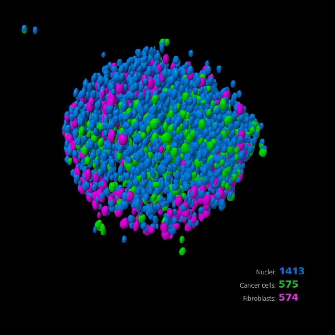
Cell Biology: Quantify with Precision
From single cells to comprehensive populations, extract meaningful data automatically
Transform your cell biology research with ZEISS arivis's powerful quantification capabilities for complex cellular phenotypes. Easily segment, track, and analyze heterogeneous cell populations in complex microenvironments that better mimic in vivo conditions. Obtain reproducible measurements that capture subtle phenotypic changes without manual intervention.
- Easily automated cell and nuclei counting with AI-based segmentation
- Cell or nuclei tracking across time series to analyze division and migration
- Confluency measurement for growth monitoring
- Phenotypic classification based on multiple parameters using AI or custom feature patterns
- Quantification of cell-cell relationships, such as distance measurements and colocalization
- Batch processing for batch and high-content experiments
While the manual workflow enabled analyzing on an average 20 cells by an individual in a day, ZEISS arivis Cloud allowed several thousands.

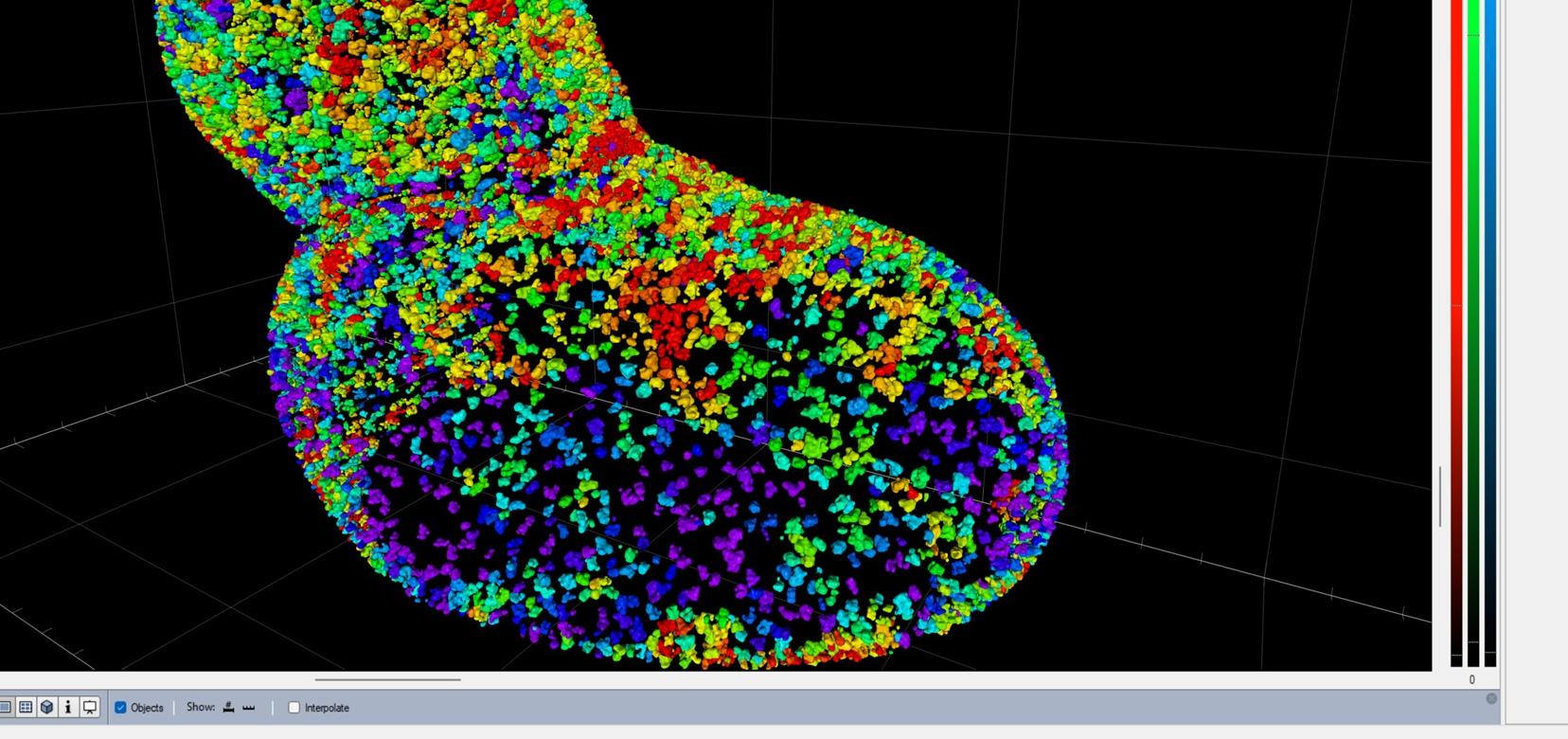
Organoid Research: Structural Analysis in 3D
From growth monitoring to detailed morphology analysis
Advance your organoid research with ZEISS arivis's comprehensive 3D analysis capabilities, supporting the transition to reduced animal testing models. Automatically segment complex structures, quantify morphological changes, and analyze cellular organization within organoids in true 3D (not just maximum intensity projections). The software's ability to handle large volumetric datasets makes it ideal for lightsheet microscopy and other 3D imaging techniques essential for more efficient translation to human patient outcomes.
- Automated organoid and lumen segmentation for volume quantification
- Cell and layer analysis within organoid structures
- Volume and feature analysis across time points to monitor development
- Robust analysis of different organoid types (intestinal, brain, kidney) and spheroids
- Phenotypic classification based on custom feature patterns or AI
- Compatibility with all major 3D imaging techniques
- True 3D analysis beyond maximum intensity projections for accurate morphology
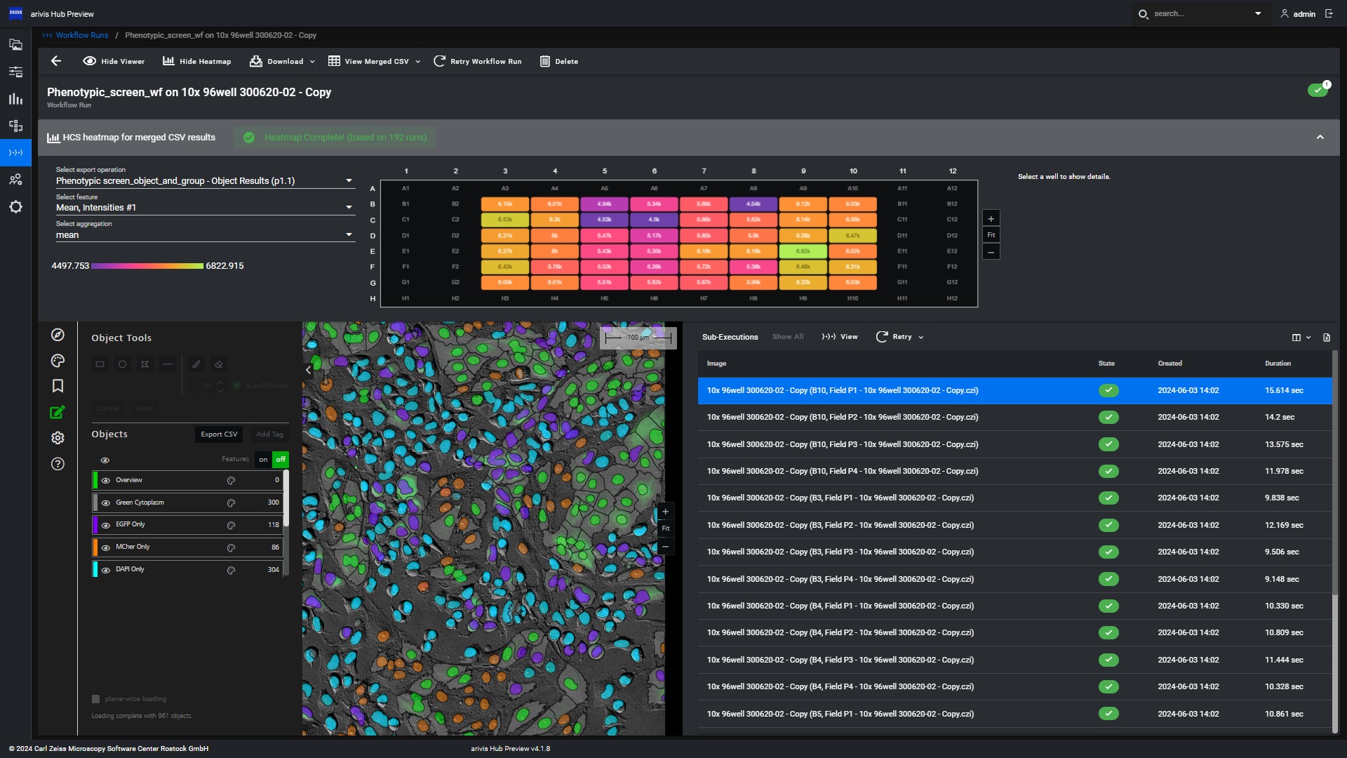
Drug Discovery & High Content Analysis: Scale with Confidence
Process, analyze, and share large datasets efficiently
Accelerate your drug discovery pipeline with ZEISS arivis's scalability for data-intensive applications. By centralizing data management and enabling parallel processing, the platform dramatically reduces time-to-results while maintaining consistency across 2D-4D assays. From individual analysis tasks to enterprise-wide deployments, ZEISS arivis adapts to support your complete drug discovery workflow with improved standardization for more reliable results and better regulatory compliance.
- Centralized data management for multiple imaging systems and file formats
- Parallelized batch processing for thousands of images
- Flexible deployment options - on-premises, cloud (AWS), or hybrid
- Automated assay workflows with folder watching capabilities to trigger execution
- Optimized resource allocation to control computing costs
- Web-based access for seamless collaboration and result sharing
ZEISS arivis Hub gives us a unique integration of all the scientific imaging data from our microscopes. The platform allows us to manage data storage, access, and processing across our facility, freeing up valuable time of our scientists to concentrate on their research questions.
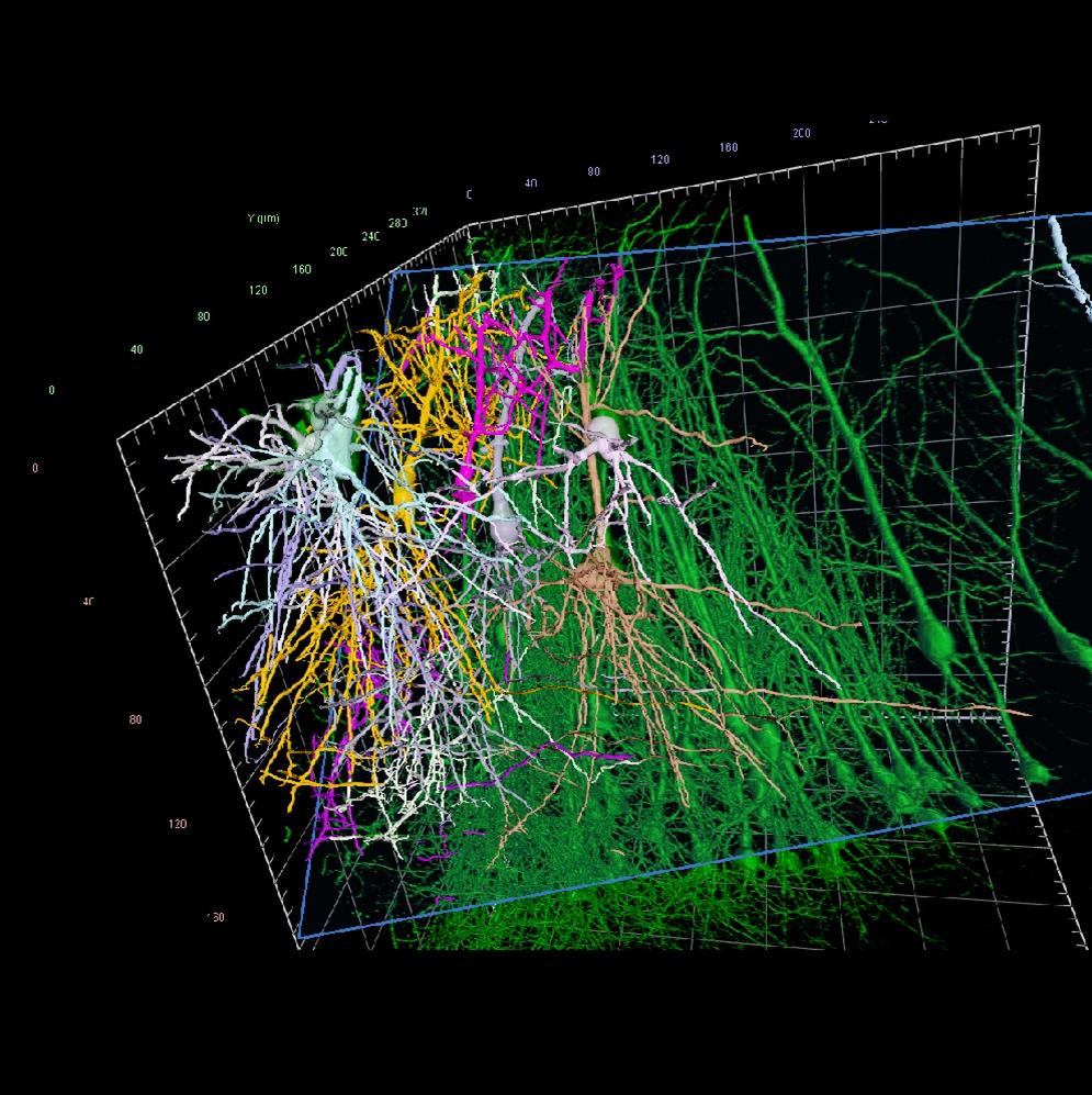

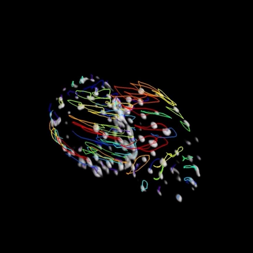
Neuroscience: Reveal Neural Complexity
From single neuron tracing to complete network analysis
Explore neural complexity with ZEISS arivis's powerful, automated tools designed specifically for neuroscience research. Trace and analyze complete neuron morphologies, and quantify dendritic spines - even in large, 3D datasets from cleared tissue or whole brain imaging. The software's advanced algorithms handle the intricate structures of the nervous system with precision, enabling discoveries that would be impossible with manual analysis.
- Automated 3D neuron tracing with state-of-the-art algorithms
- Dendritic spine detection based on state-of-the-art methods including or AI
- Large 3D dataset handling for whole brain and cleared tissue
- Volume fusion for combining several sub volumes
- Full integration into analysis workflows combining distance measurements and many more
- Comprehensive morphological measurements of neuronal features
Using ZEISS arivis Cloud allows us to save hundreds of hours spent looking at neurons and manually counting their synapses. The reproducibility of the model application ensures that the data is comparable between different researchers on the team and hopefully, in the future, between different teams as well.


Download the AI E-Book
Unlock the power of AI in microscopy with this free, newly expanded and enhanced E-Book. For Researchers In Life Sciences and BioPharma

In this free E-Book you will learn: