Influenza A virus (IAV) is an RNA virus that causes the flu in birds and some mammals, including humans. Human influenza virus usually refers to those subtypes that spread widely among humans: H1N1, H1N2, and H3N2 are the only known influenza A virus subtypes currently circulating among humans.
Allen Liu is an Associate Professor of Mechanical Engineering at the University of Michigan, USA.
Research Focus
Prof. Liu has a long-standing interest in mechanobiology of biological membranes, looking at how physical forces and stimuli affect biological functions from the vantage point of membrane remodeling and protein machineries at the plasma membrane that respond to these cues.
In this context, he and his team have been investigating how membrane and cytoskeleton mechanics govern membrane remodeling during endocytosis and cell migration. His lab is also interested in building synthetic cells from biological parts from the bottom-up. In this context, he and his team have been engineering mechanosensitive synthetic cells for a range of biomedical applications.
Understanding Intracellular Trafficking
Part of the lab focuses on investigating intracellular trafficking using various state of the art live cell imaging and super resolution fluorescence imaging techniques. They are particularly interested in clathrin-mediated endocytosis (CME), a process by which cells uptake proteins and other macromolecules by forming budded structures coated with clathrin on the plasma membrane. Various viruses, specifically viruses like Influenza A virus, SARS CoV-2, and HIV, can hijack CME and similar processes to gain access to the cell.
In their recent work, Prof. Liu's team investigated the role of epsin, a membrane bending protein, in enabling IAV entry via CME. It has been shown that upon IAV binding to the plasma membrane, epsin can specifically bind to cell surface ubiquitinated receptors. They showed that epsin’s interaction with proteins at the site of IAV binding initiated its membrane bending mechanism. Their findings show the ability of IAVs to hijack activity of membrane bending proteins to initiate membrane bending and receptor-mediated endocytosis for cellular entry.
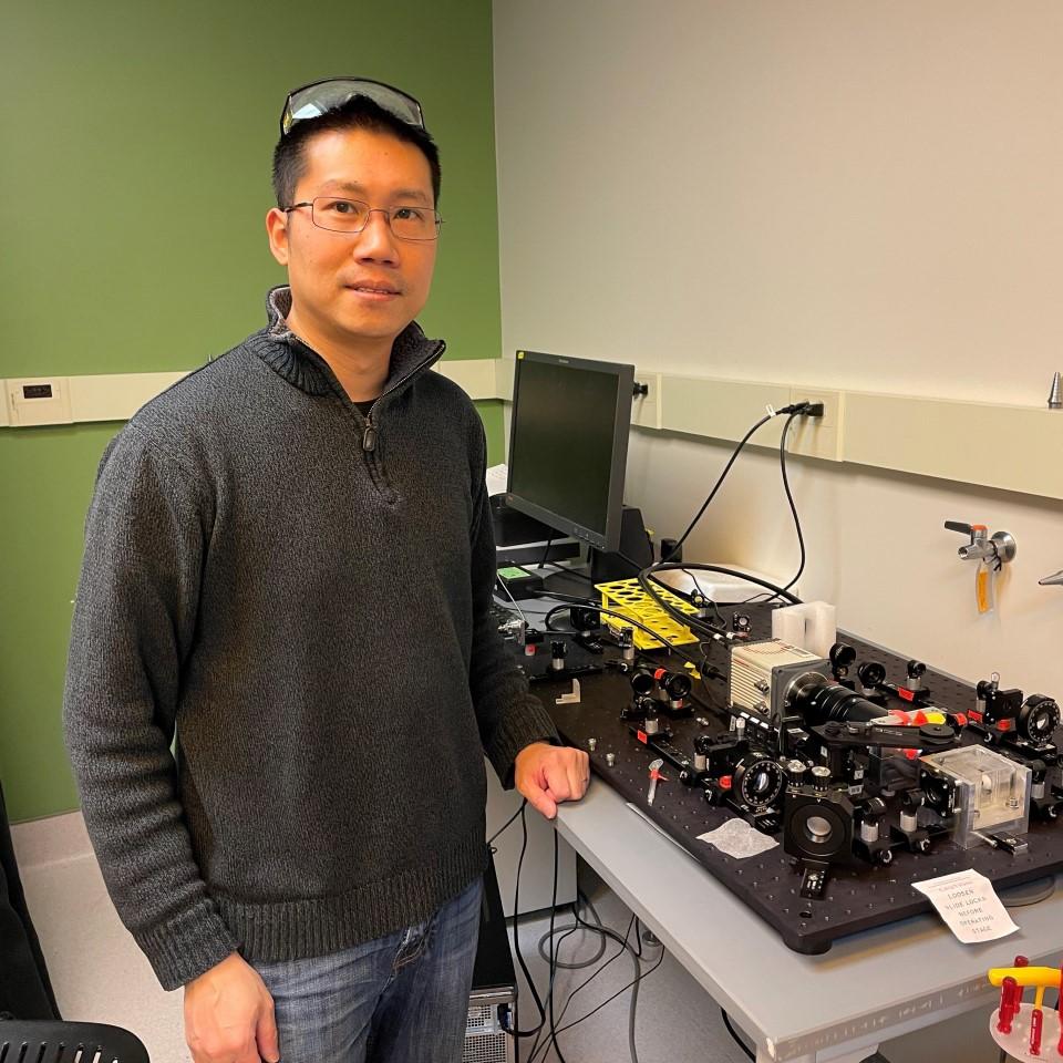
Prof Liu in front of their home-built selective plane illumination microscope set-up in the lab.
Dr Jophin Joseph, a former PhD student, significantly contributed to the research focusing on the role of epsin in IAV cellular entry in Prof. Liu's team.
Using Lattice Light Sheet Microscopy
The team utilized various high resolution live cell imaging techniques to study IAV entry in engineered cell lines with epsin and clathrin proteins tagged with fluorescent proteins. Using lattice light sheet imaging, they were able to visualize viral entry and how IAVs interact with epsin and clathrin at the plasma membrane.
Unlike traditional microscopy, lattice light sheet technique allowed us to visualize the viral entry on the entire cell surface. Furthermore, we were able to track IAVs before and after they attach to the cell membrane.
RPE cell monolayer getting infected with DiD-tagged IAV particles
This data was acquired on ZEISS Lattice Lightsheet 7 at the University of Michigan BRCF Microscopy Core Facility with the assistance of the core facility team.
Third-party Content Blocked
The video player is blocked due to your cookie preferences. To change the settings and play the video, please click the button below and consent to use of "Functional" tracking technologies.
RPE cell monolayer getting infected with DiD-tagged IAV particles, imaged with ZEISS Lattice Lightsheet 7. Green - Epsin EGFP; Red - mCherry CLC; Magenta - IAV tagged with DiD. Each frame was captured at an interval of 30 s (movies came from a Z series that took ~30 s to acquire).
Since the technique utilizes lattice light sheets which excite a thin region of the cell, they could reduce the background noise created by out-of-focus excited fluorescence proteins. This allowed the generation of high-quality viral particle tracks from which they could determine the amount of epsin and clathrin recruitment associated with the viruses. Light sheet microscopy also reduces the amount of photobleaching which enables the continuous imaging of cells for a longer period without compromising the signal quality.