

Introduction
How do the nerve cells in our brain communicate with each other? What processes take place when T cells kill cancer cells? Details of the mechanisms at the cellular level remain hidden from view. Now, special reporter proteins developed by a research team led by the Technical University of Munich (TUM) may help unveil these mechanisms. Prof. Gil Gregor Westmeyer and his colleagues at the TUM have developed a so-called genetic reporter system that does the reconnaissance work within the cells for them. The gene reporters are protein capsules just large enough to be resolved by an electron microscope.
Current electron microscope image series are a bit like looking at a city map. It is sufficient to get a visual impression of the surroundings and see where the roads are. But it doesn't tell us how often traffic lights are switched, how much traffic there is at any given point, and when or where something is currently under reconstruction.


Gil Gregor Westmeyer is a Professor of Neurobiological Engineering at the TUM and the Director of the Institute for Synthetic Biomedicine at Helmholtz Munich.
Research Focus
The work of Professor Westmeyer’s laboratory focuses on bioengineering of next-generation molecular sensors and actuators for functional imaging and remote spatiotemporal control of cellular processes. To this end, mammalian cell engineering, nanotechnology, and synthetic biology techniques are combined with multiscale molecular imaging ranging from correlative electron microscopy to magnetic resonance imaging. The new molecular imaging agents and actuators are applied for dynamic analyses of organoids and neurobehavioral imaging of preclinical model organisms to dissect cellular network function and contribute to future imaging-controlled tissue engineering as well as regenerative and cell therapies.
Identification By Barcodes
The capsules are produced by the cells themselves. Their genetic blueprints are attached to specific target genes. The reporter proteins are produced when the target genes become active. The basic principle behind this method is already a standard procedure in light microscopy. There, researchers work with fluorescent proteins. However, this method is not suitable for electron microscopy, because rather than colors, different shapes are distinguished based on their electron densities, for example.
The researchers exploited this by incorporating metal-binding proteins into differently-sized capsules. These "EMcapsulins" appear as concentric rings of various diameters under the electron microscope and can be quickly identified and assigned like barcodes using machine learning.
Labeling Neurons
By labeling neurons with EMcapsulin barcodes, it is possible to identify cellular states that would otherwise not be visible under an electron microscope. Similar to the way colors are distinguished in fluorescence microscopy, the barcodes for electron microscopy can be distinguished due to the different sizes and combinations of concentric rings.


How to Utilize These Reporter Proteins?
On the one hand, the researchers can use them to indicate the activity of certain genes, but also to localize biomolecules that would otherwise not be visible under an electron microscope – e.g. electrical synapses between nerve cells or receptors that influence the interaction between cancer cells and T cells.
The EMcapsulins can also be given fluorescent properties, which allows the researchers first to examine the dynamics of cellular processes in living tissue using light microscopy and then follow up with electron microscopy to resolve the underlying ultrastructure.
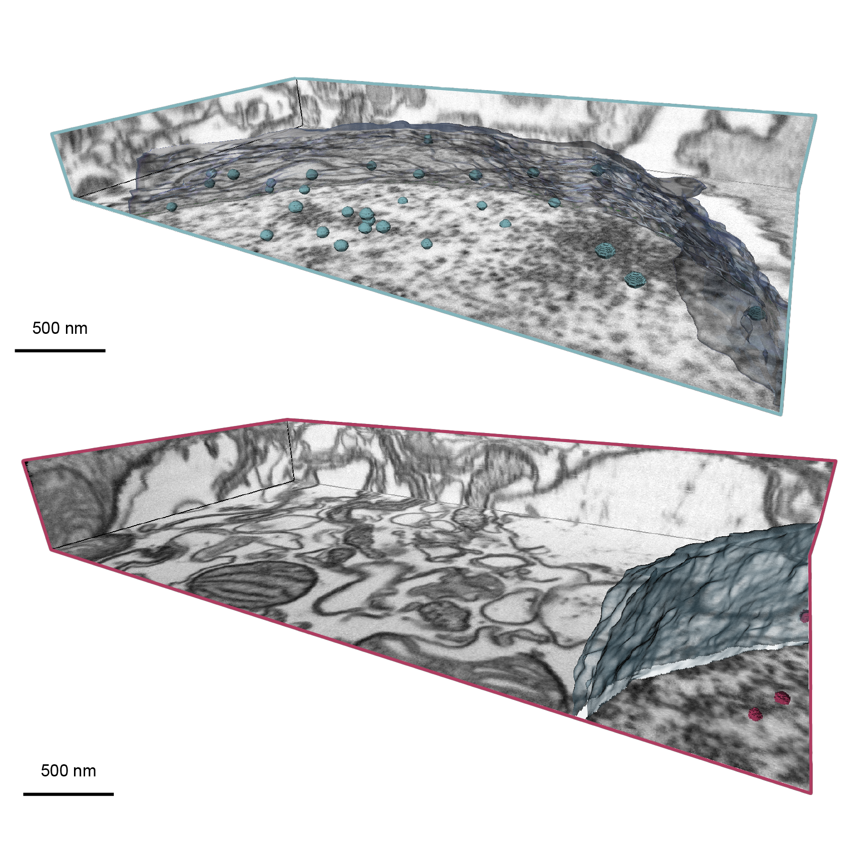

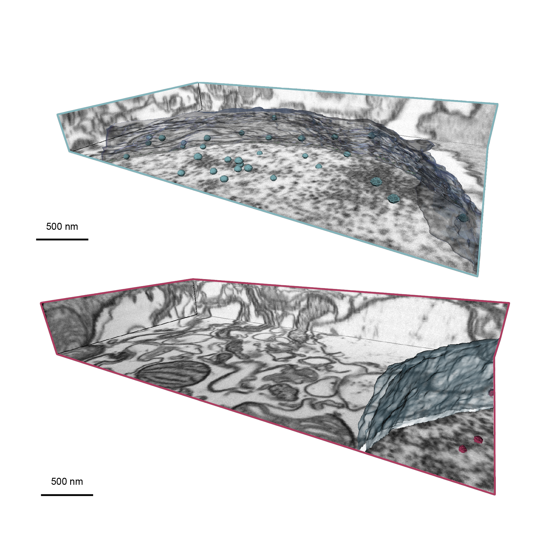

EM Imaging of EMcapsulins in Brains of Model Organisms
These Volume EM data have been acquired with Focused Ion Beam (FIB)-SEM (ZEISS Crossbeam 550). It shows Drosophila brains expressing two distinct EMcapsulins classes both targeted to the cell nucleus (segmented in blue and red).
The following settings were used for the image acquisition: SEM beam voltage 1.3 kV, a working distance of 5 mm, 6 µs target dwell time and 4 nm nominal voxel size using an InLens detector. The FIB Ga beam was accelerated by 30 kV voltage at a current of 700 pA. An image volume of 3,696 nm (width) × 1,956 nm (height) × 404 nm (milling length) was acquired in ~70 min using ZEISS ATLAS 3D software.
This figure has been adapted from Figures 5 of [1] Sigmund,F. et al. (2023). Genetically encoded barcodes for correlative volume electron microscopy. Nature biotechnology. DOI: 10.1038/s41587-023-01713-y.
It is also conceivable that, in the future, these reporter proteins could be used as sensors that change their structure, for example, when a cell becomes active. In this way, the relationships between cell function and cell structure can be better elucidated, which is also pertinent to understanding disease processes, as well as to produce therapeutic cells and tissues.
-
1
The researchers will also use the new Electron Microscopy Facility at the Technical University of Munich (TUM) and collaborate with the new TUM Center for Organoid Systems (COS).


