Expanding 3D Nondestructive X-ray Microscopy Through Laboratory Diffraction Contrast Tomography (LabDCT)
A Powerful New Complementary Solution to EBSDAbstract
Modern X-ray microscopy (XRM) combines the many remarkable characteristics of X-rays; the penetrating power and the quality to diffract off of the crystalline lattice planes (traditionally used in XRD) to interrogate interior structures of samples nondestructively providing scientists with X-ray vision via radiography or tomography within a single laboratory- based instrument. Following a trend somewhat analogous to that which was encountered previously in Scanning Electron Microscopy (SEM), wherein different electron contrast methods complemented with EDS and EBSD techniques turned the SEM from an imaging-only tool into a robust analytical platform, XRM is now expanding well beyond the classical limitations of X-ray CT or microCT. Specifically, XRM is extending the range of imaging modalities (now including phase contrast in addition to the well-known absorption contrast) and incorporating analytical diffraction information from polycrystalline samples.
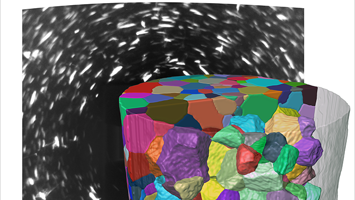
Based on a technique initially developed at a few select synchrotron facilities worldwide, diffraction contrast tomography (DCT) is now available in the lab and leverages both the absorption-based and diffraction information from X-rays’ interactions with a sample to reconstruct the crystalline microstructure in 3D. As a nondestructive method, this new analytical technique offers us the opportunity to observe phenomena which were never before possible in 3D: such as grain growth through annealing, studying the local effects of corrosion, coupling mechanical behavior with local grain structure, or correlating to complementary methods like 2D/3D EBSD. This webinar will introduce LabDCT by means of examples, and how it complements and extends the range of imaging opportunities provided by XRM, spanning from materials to life sciences.
Key Learnings:
- The basic principles and introductory information of what LabDCT is and how it works.
- How to utilize the power of extracting 3D crystallographic information non-destructively from a sample using X-ray microscopy.
- How the LabDCT technique can be applied to study various different materials shown through several applications examples.
- How LabDCT enables 4D/in-situ imaging of materials providing comprehensive microstructural and crystallographic information.
Who should watch this webinar:
Materials Scientists, engineers, and researchers working in either the academic or industrial environments interested in understanding the microstructural and crystallographic information of (single/poly)-crystalline samples.
Researchers with prior EBSD, X-ray tomography background will particularly find this webinar interesting and informative.