Measuring Communication Between the Brain and the Immune System
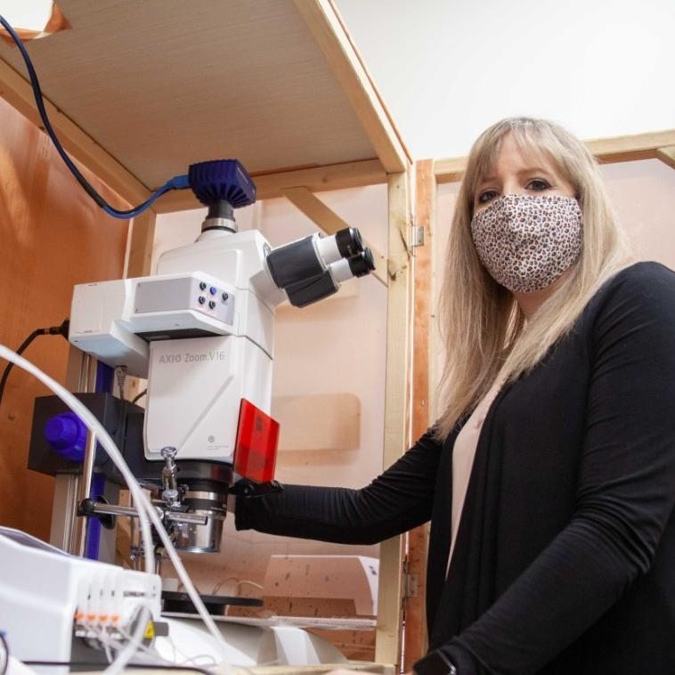
Stereo zoom microscopy used for accurate probe placement, tissue labeling and assessment
Researchers across scientific disciplines are uncovering a network of complex communication between the nervous, endocrine, and immune systems. Deciphering this bidirectional communication and its intricacies could help better understand disease pathophysiology and improve health and treatment outcomes in patients.
Dr. Ashley Ross is an Assistant Professor at the Chemistry Department and Neuroscience Program at the University of Cincinnati, USA. Her lab works on developing analytical methods to probe neurochemical signaling in the brain and immune system. Her work relies on accurate visualization and imaging provided by stereo zoom microscopy.
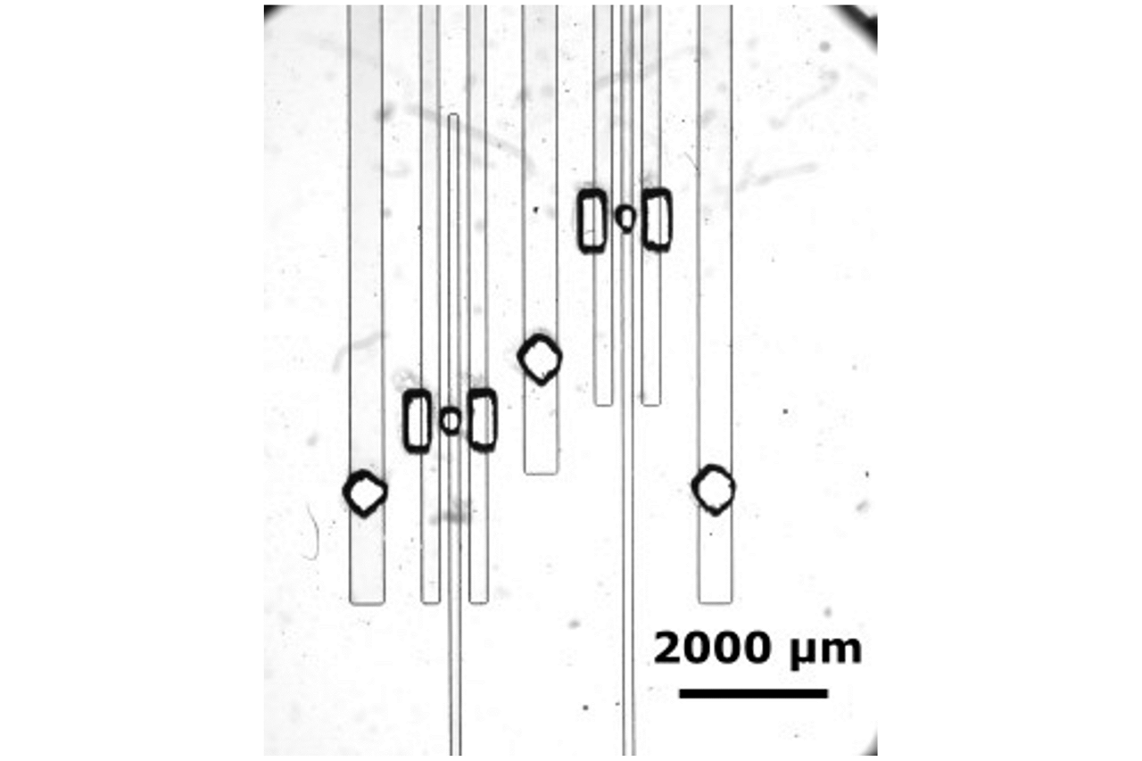
Brightfield image of a microfluidic device developed in the Ross lab to locally stimulate live tissue slices over time. The series of microchannels and ports were strategically designed to facilitate sustained localized delivery of stimuli to tissue. Imaged using ZEISS Axio Zoom.V16.
Developing New Electrochemical and Microfluidic Methods
Dr. Ross and her lab are working to develop and apply new electrochemical and microfluidic methods to measure brain-immune communication. They focus on understanding neuro-immune communication during inflammation both in the brain, directly in the peripheral immune system, and along the gut-brain axis. They have developed methods to study neurotransmitters and signaling molecules such as melatonin, guanosine, zinc, and ATP, with sub-second temporal resolution directly in tissue.
Understanding important signaling cascades during inflammation could provide critical information for developing neurochemical-targeted therapeutics in the future for immune system dysregulation.
Combining microscopy with electrochemistry is critical for us to know where we are measuring from and for assessing the health of the tissue.
Microscopy to Visualize Live Lymph Nodes and Brain Tissues
The Ross lab uses fluorescence microscopy for a variety of applications from labeling specific subregions of the lymph node, live tissue staining to accurate placement of electrodes in the tissue.
When implanting electrodes to make neurotransmitter measurements, accurate visualization is critical for exact placement within specific lymph node subregions. With live cell-surface labelling of specific "zones" in the lymph node, they can accurately place their probes in the right location for their experiment.
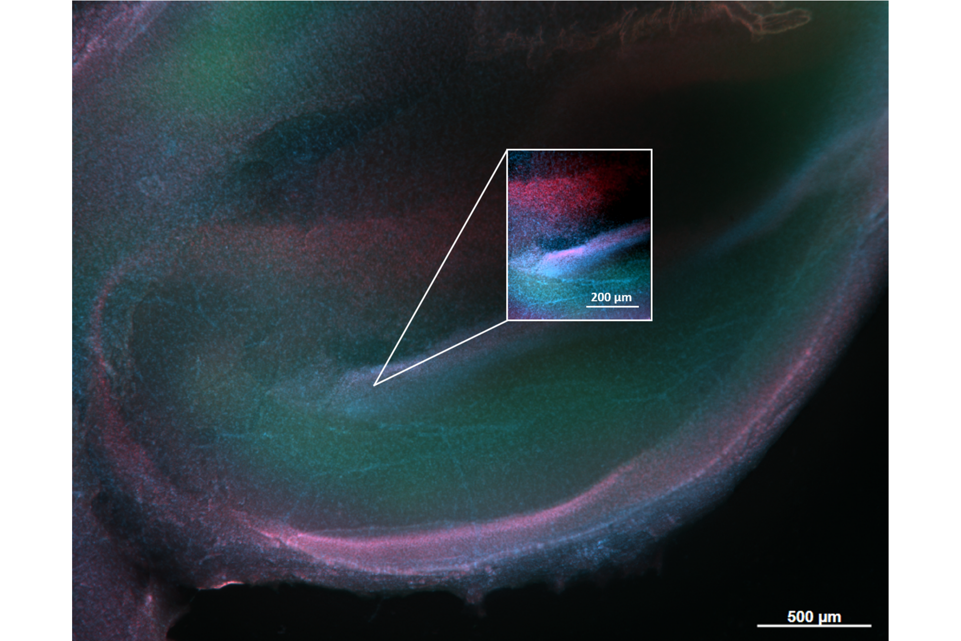
Fixed tissue fluorescence microscopy has been used to measure the "state-of-the-tissue" in post-experimentation. In one particular application, stereo zoom microscopy was used to visualize the infarct region after making measurements of guanosine signaling during focal ischemia.
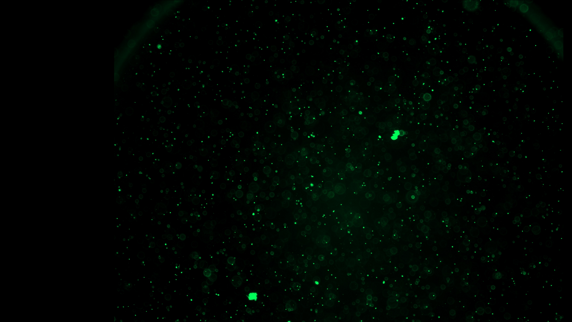
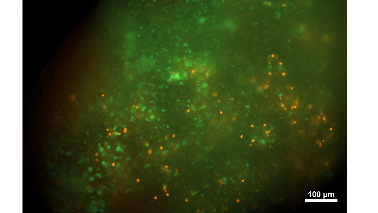
A live slice of murine mesenteric lymph node. Mice are genetically modified to express channelrhodopsin-2 (ChR2) in tyrosine hydroxylase (TH) positive neurons (genetic engineering performed in Prof. Steve Davidson Lab at University of Cincinnati, USA). The ChR2 is tagged with EYFP fusion protein for visualization (green). Slices were counter-stained with anti-TH (red) to visualize immune cells capable of synthesizing catecholamines. The Ross lab makes measurements of dynamic neurochemical signaling in the immune system to investigate the mechanism of neuro-immune communication. Imaged using ZEISS Axio Zoom.V16.
Catecholamine Fluctuations in Mesenteric Lymph Nodes
One example of their work is G.N. Lim et al., where they describe the first measurements of sub-second catecholamine fluctuations in the mesenteric (gut) lymph nodes using fast-scan cyclic voltammetry. The mesenteric lymph nodes are important for regulating gut health, which is an important piece of the signaling cascade along the gut-brain axis. Despite this knowledge, measuring neurochemical signaling directly in live lymph node, in real-time, was unexplored due to the lack of tools with adequate temporal resolution. Here, they have used fast-scan cyclic voltammetry, an electroanalytical technique often used in the brain, to make sub-second measurements in the lymph node.
This is an important advancement because it provides a tool to investigate rapid neurochemical-regulated immunity which will have far-reaching impacts on our understanding of inflammation.