Introduction
Using a ZEISS microscope he built mainly from used parts he acquired over the years, Nathan Renfro has created a beautiful gallery of microscope images of gems, rocks and minerals on his Instagram account @microworldofgems. When he isn’t in front of his personal microscope, Nathan works as a gemologist for the Gemological Institute of America (GIA). We asked Nathan to talk a bit about his journey to becoming a gemologist and his love for microscopy of rocks and minerals.


A Gemologist and Microscope Enthusiast
Microscopist and photomicrographer, Nathan Renfro with his vintage blue ZEISS Universal microscope equipped for episcopic differential interference contrast. Photo by Holly Renfro
How did you get interested in gemology?
When I was eighteen, I inherited a substantial rock and mineral collection from my grandfather who was an avid “rock hound.” I was studying at Appalachian State University in Boone, NC, USA, and would meet with the geology department chair, Dr. Jamie Allan. He helped me identify some of the many mineral specimens that were my grandfather’s. Dr. Allan eventually asked me if I would like to major in geology as I seemed to be developing an interest in minerals. I really owe him a lot for helping to point my career in the right direction.
During summer months, I worked at a local tourist gem mine called Emerald Village. While working there, I taught myself how to cut gemstones, which quickly became a new hobby. That resulted in me browsing through a copy of Lapidary Journal, a publication for gem and jewelry enthusiasts. In that journal was an advertisement for the Gemological Institute of America (GIA), where I learned it was possible to become a gemologist as a career. In that moment, I decided that’s exactly what I would do after I finished my undergraduate studies in geology. After completing their gemology program, I applied for a job at GIA and was hired as a diamond grader, transferring after a year into the Colored Stone department.
Today I am the manager of the Colored Stone department for the GIA Carlsbad and New York laboratories.


Mineral Specimen Collection
Shown here are few mineral specimens Nathan Renfro inherited from his grandfather that started his interest in gems and minerals. Photo by Nathan Renfro
What is it like working as a gemologist for GIA?
We provide a service to the gem and jewelry industry by issuing unbiased independent third party reports on gemstones. Producing this report generally involves identifying what type of stone a submitted gem is, and would also include making determinations about the natural or synthetic origin of the gem, if it was artificially improved by any sort of treatment and potentially identifying the geographic origin of the gem material.
On any given day at GIA, I could be working with something as ordinary as identifying a piece of glass that someone mistakenly thought was a natural diamond or I might examine a large, top quality sapphire from the iconic Kashmir deposit in India worth hundreds of thousands of dollars. In that regard, the variety of gemstones that I might examine daily is anything but ordinary.
When I’m not working on client stones, I’m generally staring down the oculars of my microscope or working on a research project that may involve characterizing a new gem material, like the recent one I worked on about a new deposit of pink gem diaspore from Afghanistan. Or, you might find me writing up some microscopic observation for publication in GIA’s journal, Gems and Gemology, where I am a section editor for the Microworld column.
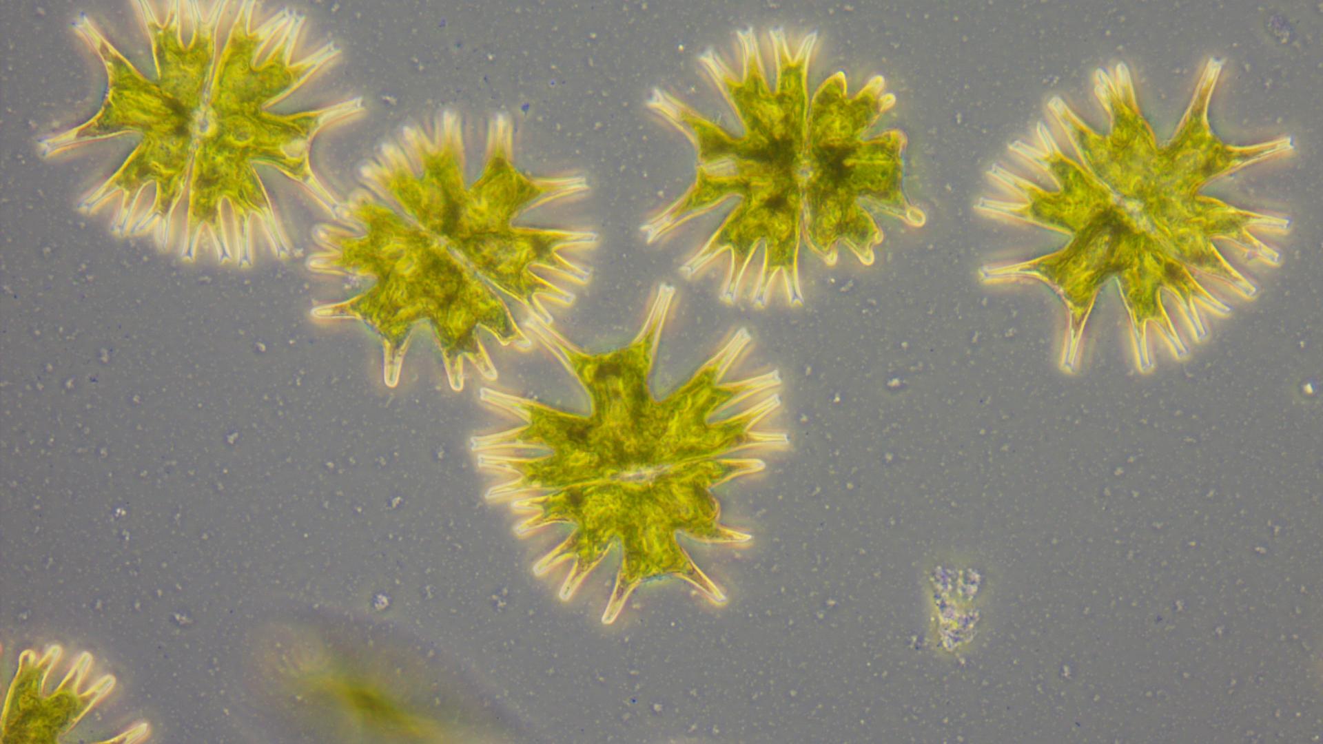
Gemstones Under the Microscope


The prism face of this natural beryl crystal shows interesting etching that resembled a mountain range. Field of View: 1.44mm.
Copyright: Nathan Renfro


This natural rock crystal quartz from Arkansas, mined by Avant Mining, showed peculiar mineral casts that are likely the result of small grains of the isometric mineral pyrite that formed on the surface in the later stage of growth of the quartz.


This synthetic corundum crystal showed a defect on the surface caused by a microscopic crystal of platinum contamination from the crucible containing the growth material.


The surface of this diamond crystal from Guyana shows the isometric crystal structure of the octahedral crystal faces as triangular etching known as “trigons” using episcopic DIC microscopy.
Field of View: 0.72mm. Copyright: Nathan RenfroWhat are your biggest challenges as a gemologist?
There are always new obstacles in the realm of new gem deposits being discovered and new treatments being developed which aren’t always disclosed. Part of GIA’s mission is to protect the public trust, so we are constantly on the lookout for new treatments that could potentially defraud the public by a lack of disclosure.
Historically, these problems can be really significant in the gem trade as was the case in 2002, when a new treatment was detected by laboratory gemologists where sapphires were diffused with beryllium in a high temperature heating process in order to improve their color. Although these things can be challenging, it keeps things interesting.
How do you use microscopy?
Gemology is primarily an observational science. Due to the high value of the gems we study, we rely on non-destructive testing to measure the physical properties of unknown gem materials. Additionally, we also use microscopy to examine gems for diagnostic inclusions, which can provide information about what a gem material is, if it has been treated in some way, and potentially the geographic origin of that particular gem.
Generally, gemologists would use a lower power stereo microscope for the types of samples we examine. Gems would be considered large samples by most microscopy standards, so in order to have any significant depth of field a stereo microscope is the way to go. As for lighting, gemologists would use a wide variety of lighting conditions including brightfield, darkfield, polarized light, and oblique fiber optic illumination, the latter of which is probably the most versatile and useful when examining gemstones.
Some years ago I was also introduced to a much more specialized microscopy method by my good friend and microscopy mentor John Koivula, who is a highly accomplished photomicrographer and microscopist. That method is episcopic differential interference contrast, or Epi-DIC, which has much more limited application, but is useful for carefully examining the surfaces of gemstones which can be used to detect treatments in some cases. Aside from the practical uses for Epi-DIC, I really enjoy the aesthetics of the bright colors and geometric forms I see on the surfaces of etched gem minerals that this microscopy technique provides.
Are there any particular samples you really enjoy imaging?
My favorite type of samples are diamond surfaces. Diamonds are a metastable mineral, which means they aren’t particularly happy at the conditions found at the surface of the earth. As diamonds make their way to the earth’s surface, the pressure and temperature decreases, causing them to begin to dissolve. This reveals intricate patterns often characterized by triangular etch features known in the gem world as “trigons”. These are always crystallographically aligned to the host diamond so all of the triangles you find on a crystal face are precisely arranged in the same orientation. Also, as diamond is the hardest natural substance, the surfaces of gem diamonds are often very pristine and undamaged providing very sharp lines and high amounts of detail of the natural morphology of the host crystal. When a diamond crystal is combined with the false color imaging technique of Epi-DIC, I’m able to produce some really interesting, colorful, abstract images that are even more special because they are of a completely natural object.


Anything else you would like to mention?
In my opinion, the digital age has allowed photomicrographers a tremendous advantage over the film days of photomicrography. Post processing, while once may have been perceived as “cheating,” is really necessary in order to resolve the most information from a digital photomicrograph. Until I learned to make use of post processing software, I always felt as though my images never looked as good on the screen as they did in the microscope. Post processing has allowed me to bring the detail back into the images so that the details they contain look as close to what I observe in the microscope as possible.
Well, this wouldn’t be a proper microscopy interview if I didn’t talk a little bit about my ZEISS microscope. In addition to using stereomicroscopes, the microscope I’m most fond of is my very vintage ZEISS Universal compound microscope that I slowly assembled over the course of a few years from parts acquired on the secondhand market, specifically for the purpose of Epi-DIC microscopy. It has a rotating stage with x/y movement which allows me a lot of freedom in composing my image as I can position my sample in any arrangement relative to the shear direction of the Nomarski prism by rotation and any position relative to the camera sensor using the x/y controls. I was also able to find a set of ZEISS epi objectives in magnifications of 4x, 8x, 16x, 40x and 100x each matched with an Inko series Nomarski prism. Because of the age of this instrument (approximately 60 years old), some of the prisms I bought had delaminated so I carefully disassembled each one, cleaned off the old cement residue and re-glued with modern optical adhesive resulting in DIC prisms that are like new. That was probably the most challenging part of this microscope project.


Nathan Renfro’s Vintage ZEISS Universal Microscope
The other detail that is probably apparent with my microscope is that it is now bright blue. Blue is my favorite color, so I wanted to put a modern spin on the classic look of this instrument. I left the black portions their original color for contrast, but disassembled the body and painted each piece before putting everything back together. I even painted the power supply to match. I have always thought of the design of the ZEISS Universal microscope to be a beautiful work of art on its own with its dramatic, curving spine, shiny aluminum knobs and trim pieces accented with bold black components like the stage, optovar and head. I wanted my version to be a modern take on that, which is why I decided to paint it a fun color. Every time I go to my office and see my blue ZEISS, it just makes me happy.
In Brief
-
Gemology is the study of gemstones, including their identification, grading, and valuation. Gemologists analyze the physical and optical properties of gems to determine their authenticity and quality.
-
Microscopy is crucial in gemology for non-destructive testing and examining gemstones for diagnostic inclusions. It helps gemologists identify the type of stone, determine if it has been treated, and ascertain its geographic origin.
-
In gemology, microscopes are essential tools for examining the internal and external characteristics of gemstones. Stereo microscopy is particularly important for examining the surface and internal features of a gemstone in great detail. Polarized microscopy is useful for distinguishing minerals, identifying inclusions and studying stress patterns in stones. Scanning electron microscopy is used in advanced gemology for high-resolution imaging of inclusions, surface texture and to study the chemical composition of gemstones. For the identification of complex internal structures and 3D reconstruction of gemstone surfaces and inclusions, laser scanning microscopy is the right choice.
-
Gemologists face challenges such as the discovery of new gem deposits and treatments that may not be disclosed. They must stay informed about these developments to protect public trust in the gem trade.