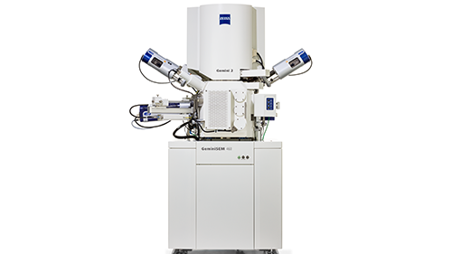
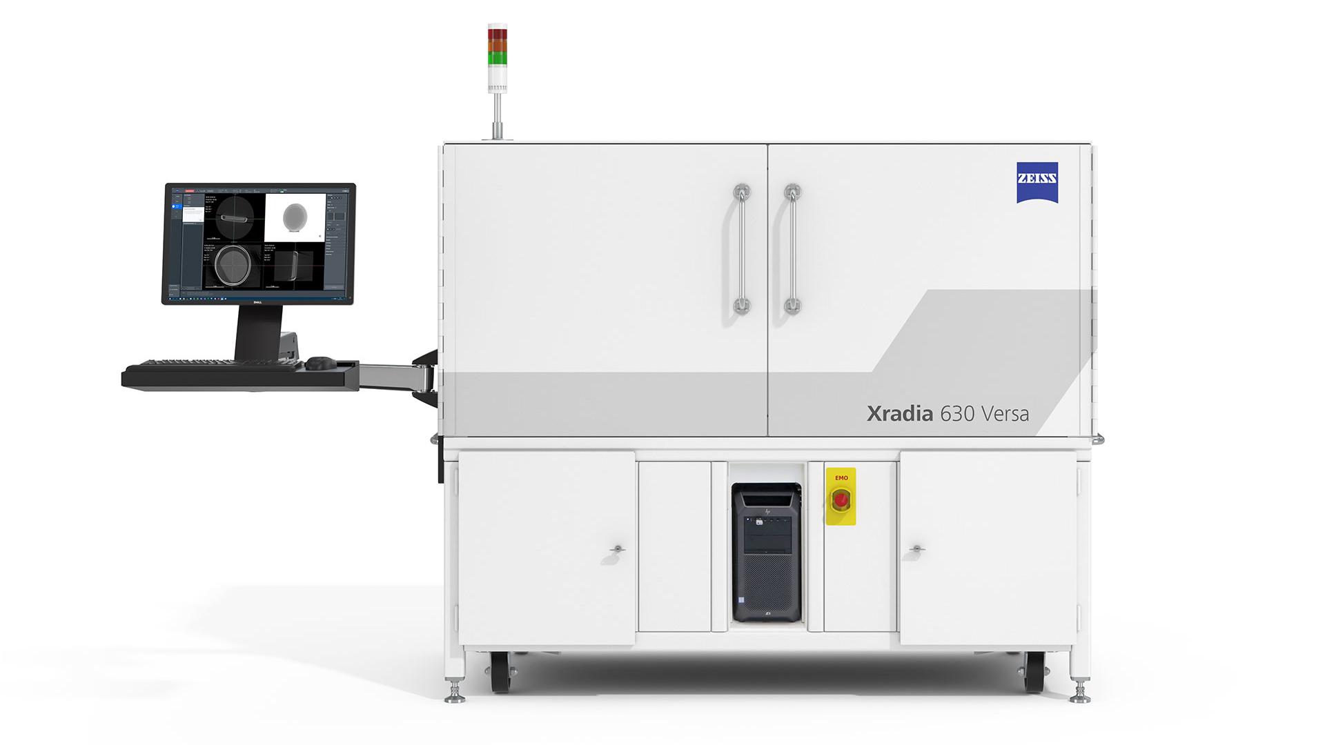
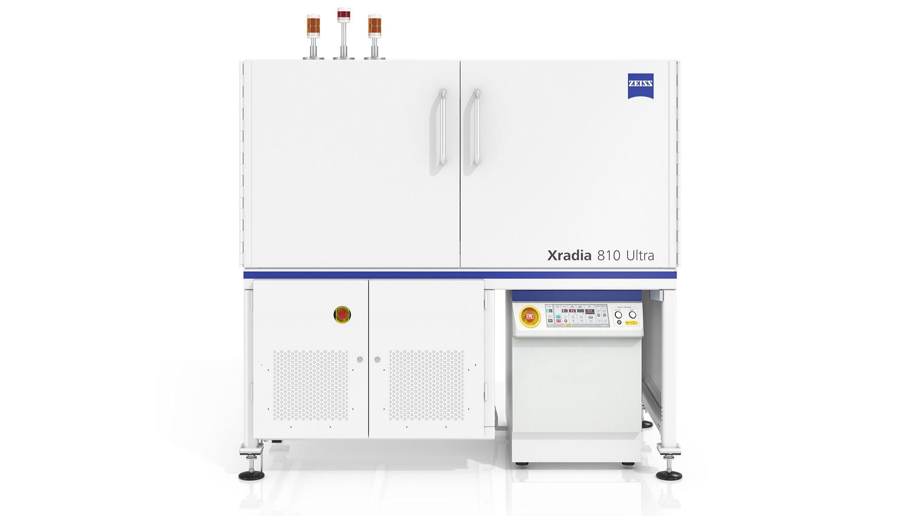
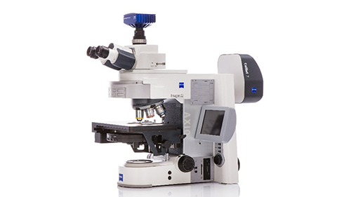
Microscopy Solutions for Materials R&D

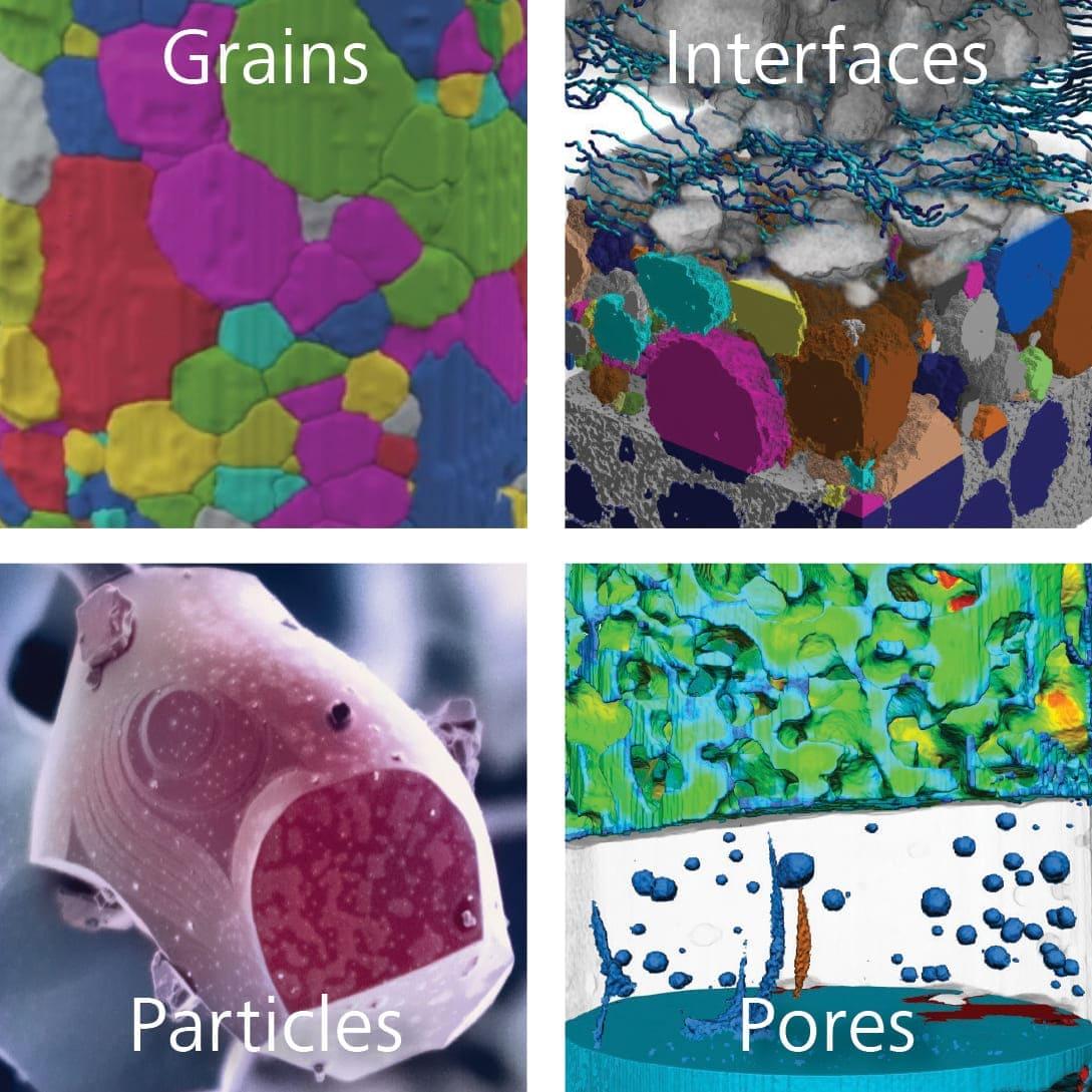
Understand the four key areas of materials research

Grains, interfaces, particles, and pores. How do we understand these properties of our materials and their influence on performance?

Using ZEISS microscopes, you can  

  • Characterize grains and crystals, particularly in metals and crystalline materials..
  • Study particles and inclusions in steel, aggregates, polymers, ceramics, catalysts, or additively manufactured parts.
  • Analyze interfaces and surfaces in batteries, electronic materials, coatings, and layers.
  • Reveal porosity, cracking, and voids and their role in macrostructural behavior.

10 Materials in 2 Minutes

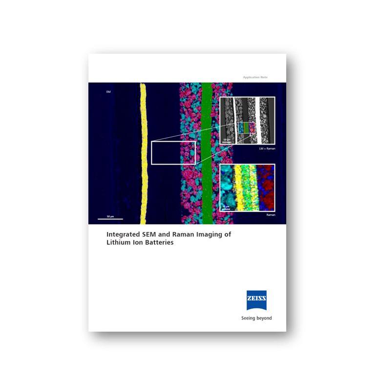
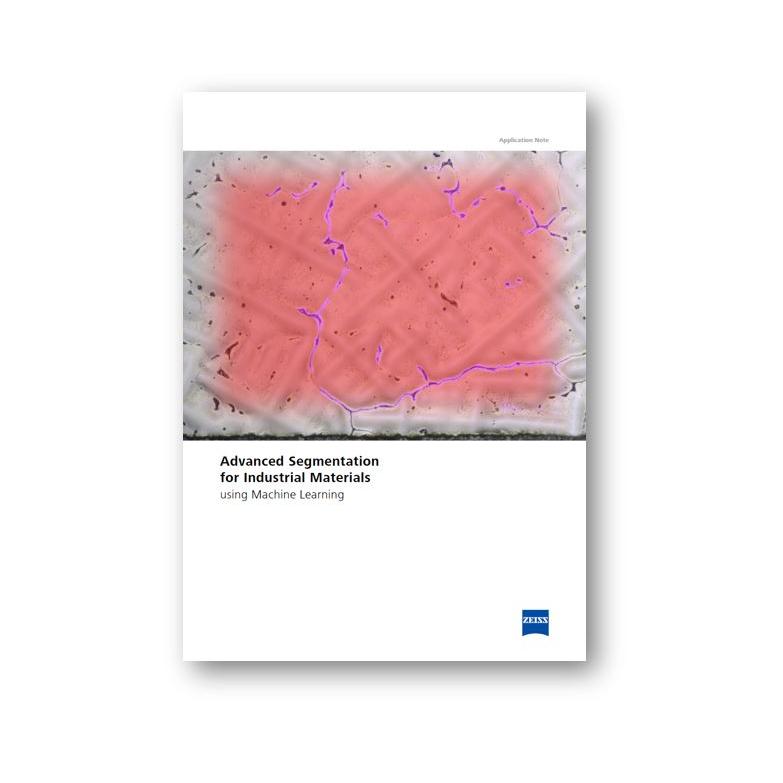
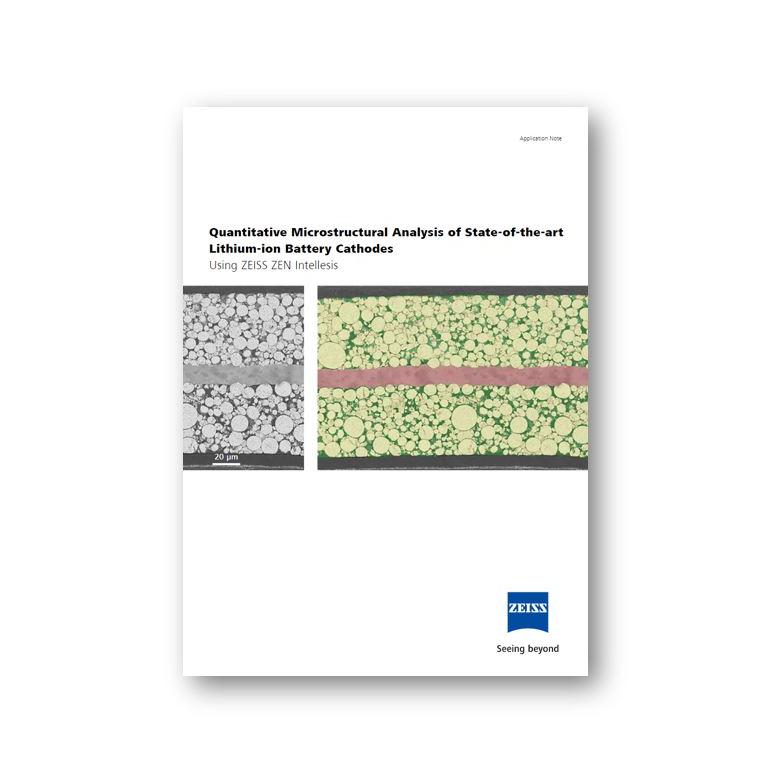
To learn more, we invite you to download these papers

  • Engineering Materials: Metals and Alloys

    New stronger, tough, durable, lightweight and fracture-resistant materials open doors to new technologies, whether it is in the applications of high-speed air or ground transportation, long lasting and economical construction, safe nuclear installation, or more advanced space exploration technologies. Designing and developing the engineering materials for tomorrow’s innovation challenges demands an ever-better understanding of the complex connections between processing, properties and the underlying structures that, collectively, influence how a material performs and why it might fail.

  • Investigating Sweet Spot Imaging of Perovskite Catalysts Bearing Exsolved Active Nanoparticles

    Platinum (Pt), generally dispersed on a solid oxide support, has been widely used for catalytic chemical reactions in automobile, chemical refining, and energy industries. During the reactions, Pt is exposed to severe conditions, e.g., high temperature events and impurities, that cause Pt agglomeration and poisoning, respectively, resulting in activity/stability losses. Perovskite materials are designed with Pt for significant catalytic properties through novel doping and exsolution methods 1. In order to accurately determine the catalytic ability of Pt nanoparticles, it is important to understand the structure and morphology of nanoparticles. Typical scanning electron microscopy methods do not reveal the morphological characteristics of nanoparticles due to the lack of electron beam stability. Here we demonstrate imaging techniques employed to accurately determine particle size and morphology. This method can improve the catalytic analysis of Pt loading, size, dispersion, and active sites determination.

  • Detection, Quantification and Advanced Characterization of Non-metallic Inclusions in Steels

    ZEISS X-ray Microscopes

    Non-metallic inclusions (NMI) are present in all grades of steels and related alloys to a greater or lesser extent and can be divided into two categories. Indigenous inclusions occur within the metal itself, as a product of chemical reactions within the melt. These reactions may occur with alloying elements, gaseous species or with impurities introduced from recycled material. Exogenous inclusions are caused by the entrapment of external non-metallic material; this may include pieces of refractory linings or dross and slag materials. Non-metallic inclusions may be oxides, sulfides, silicates, nitrides or a number of other non-metallic compounds, furthermore each inclusion may contain multiple different phases.

  • Multi-modal characterization and advanced analysis options for industry and research

    Metallography has long been the focus of the metallurgist. Routine inspection and quality control tasks can be achieved rapidly and accurately using light and electron microscopy in industrially-focused workflows; now, multi-modal microscopy is capable of providing a vast array of analytical capability, in situ and often non-destructively. ZEISS offers solutions focused on both the industrial researcher and the quality engineer around five key areas: chemistry, crystallography, dimensional measurement, tomography, and determining processing parameters.

  • Enabling Premium 3D Crystallographic Imaging in Your Laborator

    X-ray tomography for 3D non-destructive imaging has been widely adopted and operated under two primary contrast mechanisms for quite some time: X-ray absorption and phase contrast, which both rely on material density differences within the sample. However, single-phase polycrystalline materials (e.g., steels, alloys, ceramics) do not exhibit any absorption contrast that reveals the underlying grain microstructure. Synchrotron-based X-ray imaging methods – such as diffraction contrast tomography (DCT), which provide crystallographic information from the diffraction signals of single-phase polycrystalline samples, non-destructively and in three dimensions (3D) – were the first to successfully demonstrate results in this class of materials almost two decades ago. Now, advancing laboratory X-ray microscopy (XRM) one step further, we describe here the latest capabilities of laboratory-based DCT on ZEISS Xradia 620 Versa and ZEISS Xradia CrystalCT 3D X-ray imaging systems, and present the new research and 3D characterization capabilities this enables.

  • Integrated SEM and Raman Imaging of Lithium Ion Batteries

    Compared to lead-acid batteries, lithium-ion batteries (LIB) have a higher specific energy as well as higher specific power, thus they have become the most popular power source for portable computing and telecommunication equipment. Today, the main driver of battery research projects are the automotive industry, developing portable batteries, and the renewable energy market, discovering new materials for stationary energy storage as well as the electronics industry. The battery commonly consists of two electrodes that are isolated by a separator and soaked in electrolyte to promote the movement of ions. Usually graphite is the main component of the anode. On the side of the cathode mainly lithium transition metal oxides such as Li(NixCoyMnz)O2 (NCM) or LiMn2O4 (LMO) are used as active materials. To achieve specific cell properties composite electrodes with two or more different active materials are also widely used.

  • Advanced Segmentation for Industrial Materials using Machine Learning

    Since the development of the first microscope cameras, researchers and operators have sought to extract quantitative, actionable information from micrographs to further advance their research and improve their processes. Every quantitative analysis of a set of micrographs involves some form of segmentation. Segmentation is the division of images into defined regions for subsequent categorization and analysis. A region could be a mineral fragment, a grain in a metal, a pore in a composite, an oil contamination on the surface, a blood cell – any area differentiable from a neighboring area. By analysing these regions or the borders between regions, we get useful information. There are several standards for determining useful microstructural properties by image analysis of segmented images – e.g. for grain size (ASTM E112), graphite in cast iron (EN ISO 945-1), inclusion content (ASTM E45) and porosity in ceramic coatings (ASTM E2109). These analyses, however, depend on the accuracy and reliability of the segmentation results.

  • Quantitative Microstructural Analysis of State-of-the-art Lithium-ion Battery Cathodes

    In this application note two different segmentation methods – classic thresholding and machine learning- based – are evaluated in the context of quantitative analysis of constituents of state-of-the-art lithium-ion batteries. Both methods are compared against reference measurements using a laboratory balance. A detailed description of the fundamental functionality of the machine learning based approach is given. It has the potential to be more robust against image variations and thus can provide a more accurate segmentation result.

Get more information

FIB-SEM tomography dataset acquired from a commercially purchased 3D NAND sample.

ZEISS Solutions for Industrial Ceramics Research

The engineering of ceramics through characterization is about understanding the structure, properties, performance, and process of a particular material or system.

FIB-SEM tomography dataset acquired from a commercially purchased 3D NAND sample.

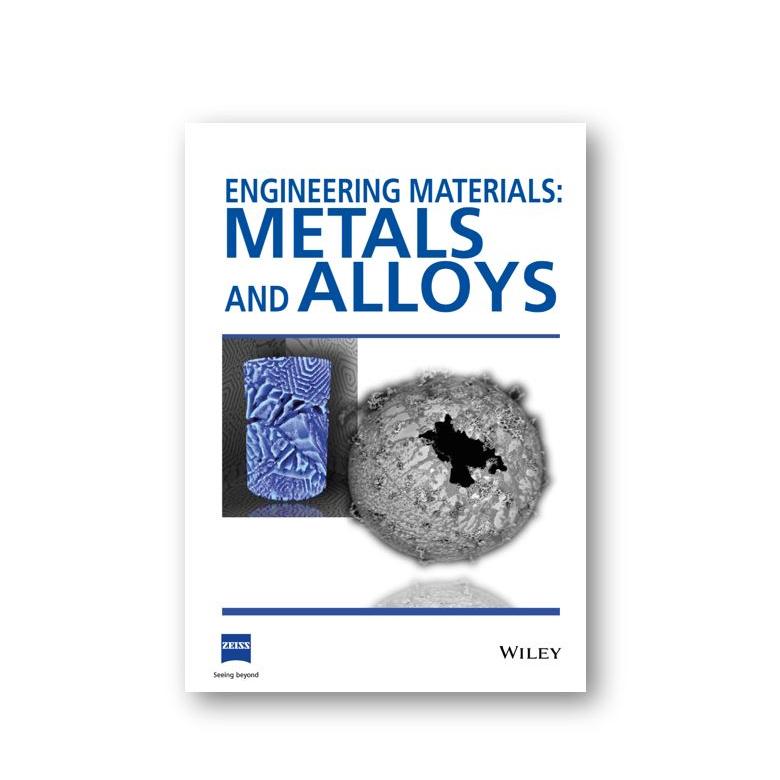
ZEISS Solutions for Metals and Alloys

Building the future with stronger, tougher, lighter and sustainable metals and alloys

FIB-SEM tomography dataset acquired from a commercially purchased 3D NAND sample.

ZEISS Imaging Solutions for Catalysts, Chemistry, Coatings and Corrosion

ZEISS imaging systems provide powerful detail for your analysis of corrosion in waterborne systems.

Download the collection here ↓

Please fill the form below for instant access to all papers

Form is loading...

For more information on data processing at ZEISS, please refer to our data privacy notice.

Related Products

With advanced light microscopy, nondestructive submicron CT to nanoscale X-ray microscopy, and high-resolution scanning electron microscopes, with AI-based solutions that simplify or speed your time to answers, ZEISS offers a full portfolio that helps you find answers to your critical development questions.


Share this Page