

Deconvolution Toolkit Methods for Computational Optical Sectioning in Life Sciences
Even the most advanced imaging systems suffer from unavoidable image blur by optical and electronic microscope components. Fortunately, this can be corrected with computational deconvolution methods. The Deconvolution Toolkit provides a collection of such algorithms for life science applications.


Improve Your Image Quality by Removing Image Blur
Solve the problem at its rootImage blur is inevitably introduced by the laws of optical physics and the use of electronic components of imaging systems. This reduces image quality metrics such as signal-to-noise ratio, image contrast, axial resolution, and lateral resolution. Applying deconvolution to your images allows you to optimize each of these image quality metrics or, optimally, all of them at once.
Image: Rat cortical primary culture; projection (extended depth of focus) of a 4-channel z-stack; GPU-based deconvolution. Sample courtesy of H. Braun, LSM Bioanalytik GmbH, Magdeburg, Germany


A Range of Algorithms Designed to Fit Your Needs
Select based on your priorities, from processing speed to uncompromised imaging qualitySeveral different deconvolution algorithms have been developed. Some methods are fast and sufficient for systems with low levels of blur. When systems present more challenging levels of blur, more sophisticated algorithms are required that use much more extensive computational resources and time. While these can solve more difficult problems and yield superior image results, the processing time can sometimes present too much of a barrier. ZEN Deconvolution Toolkit offers a range of algorithms, enabling you to choose the best fit for your experimental and post-processing needs.


Improve Images from Various Imaging Systems
Deconvolution is more than a tool for just widefield microscopyWhile deconvolution is traditionally thought of as a tool for widefield fluorescence microscopy, all imaging systems have blur resulting from optical equipment and electronic components. Whether you work with conventional widefield, Apotome, an LSM system or a Lattice Lightsheet 7, all these imaging systems can benefit from including deconvolution into your image processing workflows. For each imaging system, certain recommended algorithms are more suitable and available to use for optimal results.


Easy to Use and Optimized for High Performance Capabilities
Improved features automate your workflowThe Deconvolution Toolkit algorithms have been optimized to make it extraordinarily easy-to-use and high performing. ZEN reads the image’s metadata and automatically determines the point spread function (PSF) of the optical system to adjust the optimal settings for deconvolution. Algorithms are accelerated by more than 10-fold by GPU or even mGPU processing. To optimize time-to-results, all deconvolution algorithms run with ZEN Direct Processing during image acquisition. And the lossless data compression algorithm Zstandard (zstd) further accelerates resource-intensive processing.
Image: U2OS cells labeled for mitochondria (TOM20-mCherry) and microtubules (Tubulin-GFP) structures before and after Constrained Iterative Deconvolution.
ZEN Deconvolution Toolkit – Basics and Application
How to separate image content and image blur?")
")
The Point Spread Function (PSF) – Why noise-free images are physically impossible
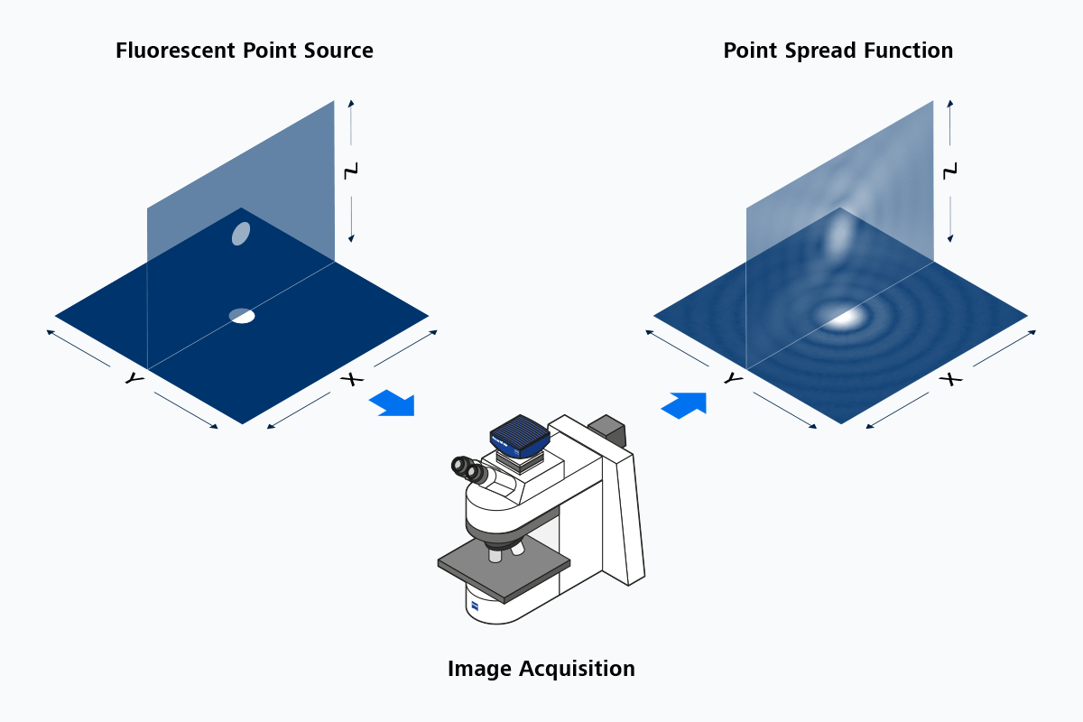

When observing any object at the scale of a single wavelength, the resulting image is inevitably blurred. Fortunately, the resulting blurring pattern, the point spread function is similar for every object. It can be directly observed when imaging a fluorescent point source.
The Point Spread Function (PSF)
Why noise-free images are physically impossibleWhen observing any object at the scale of a single wavelength, the resulting image is inevitably blurred. Fortunately, the resulting blurring pattern, the point spread function is similar for every object. It can be directly observed when imaging a fluorescent point source.


Deconvolution Principle – Applying the PSF to digital images
Convolution is a mathematical operation where one function is modulated by another. Image blurring can be understood as the convolution of the real-world objects with the PSF. Luckily, this mathematical process can be inverted, and this is what happens during deconvolution. By convolving the blurred image with the inverse of the PSF, we generate a denoised image.
What is Deconvolution?
Applying the PSF to digital imagesConvolution is a mathematical operation where one function is modulated by another. Image blurring can be understood as the convolution of the real-world objects with the PSF. Luckily, this mathematical process can be inverted, and this is what happens during deconvolution. By convolving the blurred image with the inverse of the PSF, we generate a denoised image.


Deconvolution Fine-Tuning – For advanced users
The highest quality deconvolution requires more information about the exact PSF of your system, including sample preparation. ZEN allows you to enter your own experimental PSF, fine-tune optical parameters and PSF by providing information about sample embedding medium, cover glass thickness, PSF depth variance, and many more parameters.
Deconvolution Fine-Tuning
For advanced usersThe highest quality deconvolution requires more information about the exact PSF of your system, including sample preparation. ZEN allows you to enter your own experimental PSF, fine-tune optical parameters and PSF by providing information about sample embedding medium, cover glass thickness, PSF depth variance, and many more parameters.
How do Deconvolution Methods Differ?
The basic processes of convolution and deconvolution in the context of image blurring and the PSF is the key to understand deconvolution algorithms. Every deconvolution algorithm in ZEN uses some variation of these.Simple algorithms like deblurring or nearest-neighbor subtract a PSF-generated image blur from the raw image.
The Inverse Filter method directly applies the inverse PSF. Iterative methods apply the forward PSF to approximate the raw image.
|
|
|
|
|
|
|---|---|---|---|---|
|
Deconvolution Method |
Algorithmic Process |
Application |
Speed |
Result Quality |
|
Deblurring |
Subtract blur from same image plane |
2D data sets |
very fast |
medium (2D) |
|
Nearest Neighbor |
Subtract blur from neighboring planes |
3D with limited stack |
very fast |
low |
|
Inverse Filter |
Apply inverse PSF function |
Full 3D stack |
fast |
medium |
|
Fast Iterative (Meinel) |
Iterative; Forward PSF on predicted image; Error function |
Perfectly symmetric PSFs |
medium |
good |
|
Fast Iterative (Richardson-Lucy) |
Iterative; Forward PSF; Maximum likelihood |
Asymmetric PSFs |
medium |
good |
|
Constrained Iterative |
Iterative; Forward PSF; Optimized maximum likelihood |
Gold standard |
slow |
very good |
Deconvolution Default Settings and Options
For most relevant imaging systems|
|
|
|
|
|
|---|---|---|---|---|
|
Imaging modality |
Widefield |
Apotome |
Confocal |
Lightsheet |
|
Normalization |
Auto |
Auto |
Auto |
Auto |
|
Background correction |
off |
off |
off |
off |
|
Flicker correction |
off |
off |
off |
off |
|
Decay correction |
off |
off |
off |
off |
|
Hot pixel correction |
off |
off |
off |
off |
|
Constrained Iterative specific defaults |
||||
|
Strength |
Auto |
NA |
Auto |
Manual=5 |
|
Likelihood |
Poisson |
NA |
Poisson |
Poisson |
|
Regularization |
Zero order |
NA |
First order |
Zero order |
|
Optimization |
Analytical |
NA |
Line search |
Analytical |
|
First estimate |
Input |
NA |
Mean |
Input |
|
Max interactions |
40 |
NA |
7 |
40 |
|
Auto stop percentage |
0,1 |
NA |
0,1 |
0,1 |
|
Fast Iterative specific defaults |
||||
|
Method |
Meinel |
NA |
Richardson Lucy |
Meinel |
|
Regularization |
None |
NA |
None |
None |
|
Optimization |
None |
NA |
None |
None |
|
Max Interactions |
15 |
NA |
50 |
15 |
|
Auto stop percentage |
0,1 |
NA |
0,1 |
0,1 |
|
Regularized Inverse Filter specific defaults |
||||
|
Regularization |
Zero order |
Zero order |
Zero order |
Zero order |


Cybersecurity at ZEISS Microscopy
As digitalization advances in microscopy, so do the complexities of cybersecurity. ZEISS Microscopy is committed to proactively securing our technologies and protecting our customers. Our Cybersecurity and Data Privacy Governance Program goes beyond traditional security—it also encompasses Responsible AI and Open Source Software (FOSS) governance.


