

Multi-Scale battery imaging
Applications such as electric vehicles (EVs) and grid storage are driving market growth in battery technology. But several important materials challenges need to be overcome before next-generation batteries become standard in these areas. For example, safety is of particular concern due to the flammability of certain electrolytes and the high activity of battery cells in EVs and portable electronics.
Optimizing a battery means understanding its structure on multiple scales
Future battery research will be focused on power density improvement through new electrode designs. The microstructure of these electrodes plays a crucial role in performance, such as the driving range and charging ability of EVs. And the evolution of the electrode over time determines its lifetime stability - dendrite growth and cracking can result in short circuits, causing a battery to fail prematurely or catastrophically. So bulk defects like foreign particles and dendrites mean that quality control is critical when it comes to safety.
To optimize performance for a specific application and prevent premature failure, you must study the battery on multiple scales. This includes the composition, crystal lattice structure, and the microstructure of the individual materials. It also means studying the electrode and packaging level, where you can understand the overall integrity and enclosure of the cell. And for the most accurate results, you have to perform this multi-scale analysis in situ without damaging your sample.
Non-destructive imaging of battery electrodes is crucial
Non-destructive imaging of a battery cell is needed at these different scales, but without compromising the structure. This is challenging because batteries are sensitive to air, and processing makes analysis difficult due to complex sample preparation methods. These might include cutting or opening, disassembly, and mounting.
ZEISS microscopy solutions allow you to solve these pressing research challenges in battery technology. Non-destructive X-ray microscopy for battery analysis is crucial if you want to study your samples at varying length scale without compromising the battery integrity or structure. In other words, you can take high resolution images without exposing sensitive components to air. This allows you to gain critical insights into battery lifetime changes, failure modes, and defects with in situ imaging and preserve the battery for further analysis via correlative and multiscale workflows later.
Your next step
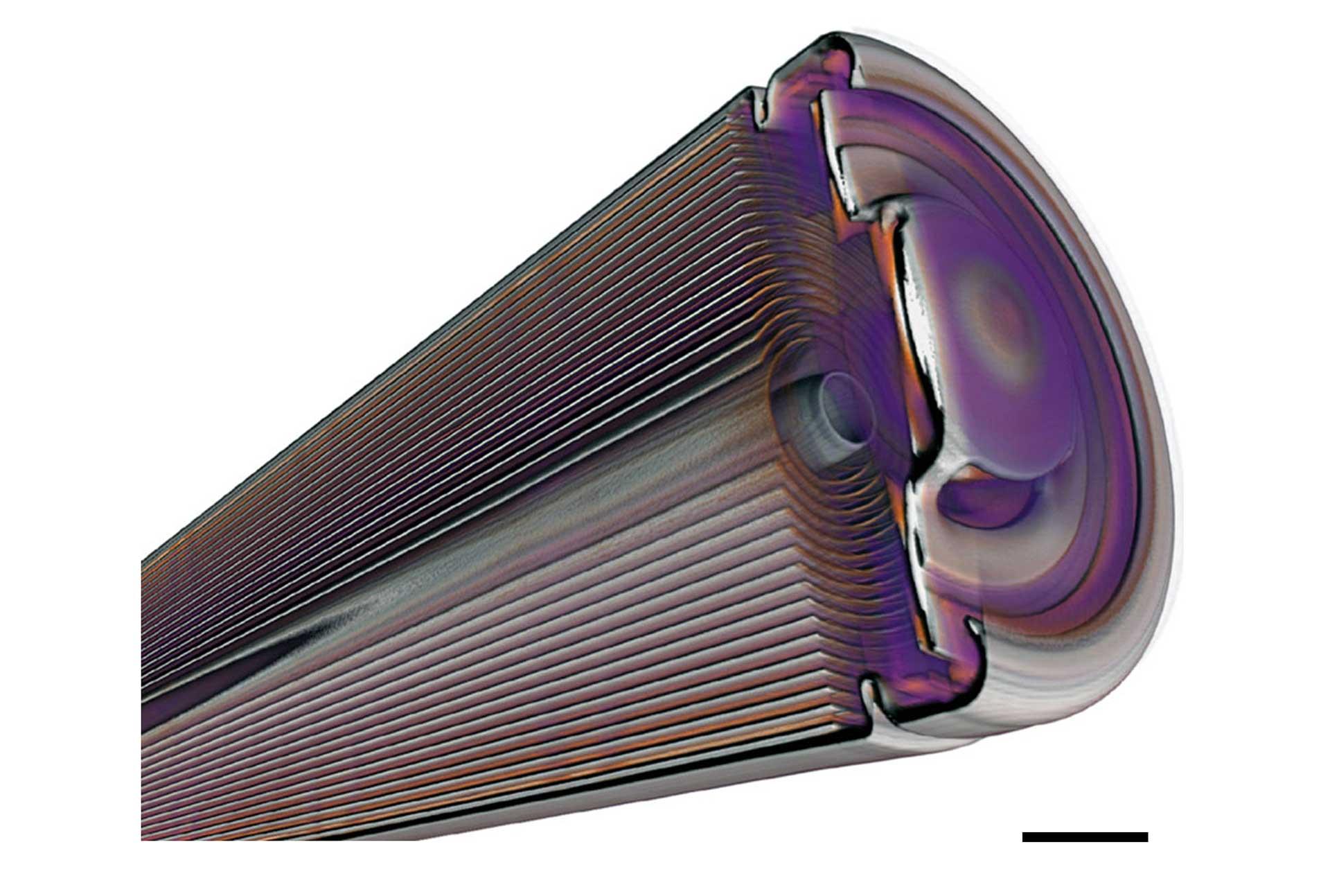
ZEISS has a comprehensive and correlative portfolio that allows you to take non-destructive images at different lengths scales in 2D, 3D and 4D.
How-to video
Application images


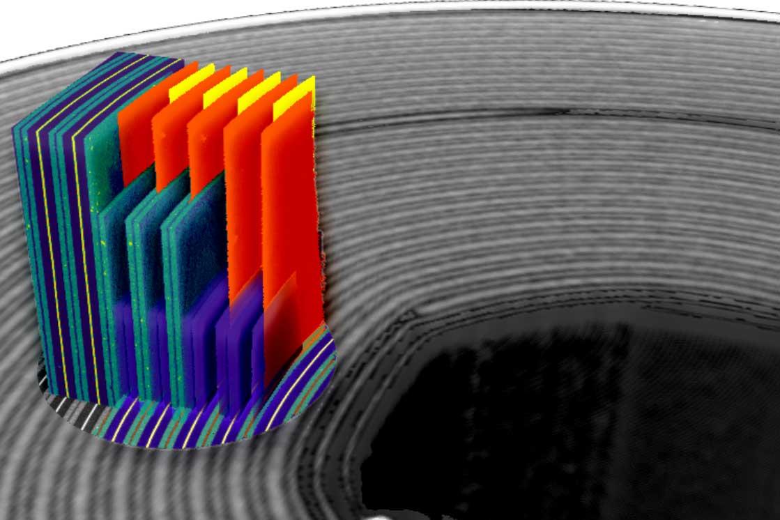

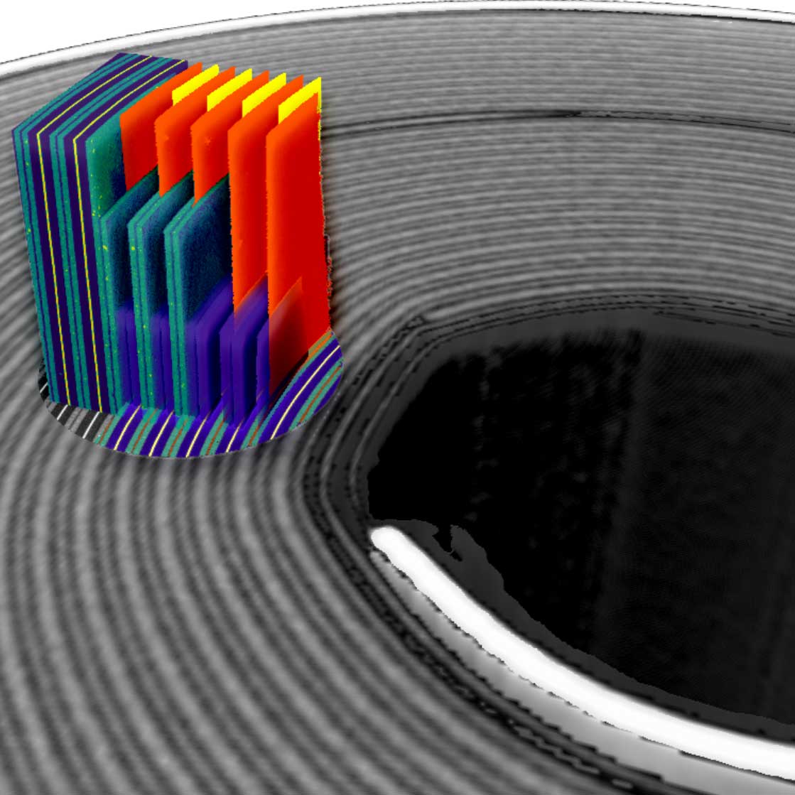


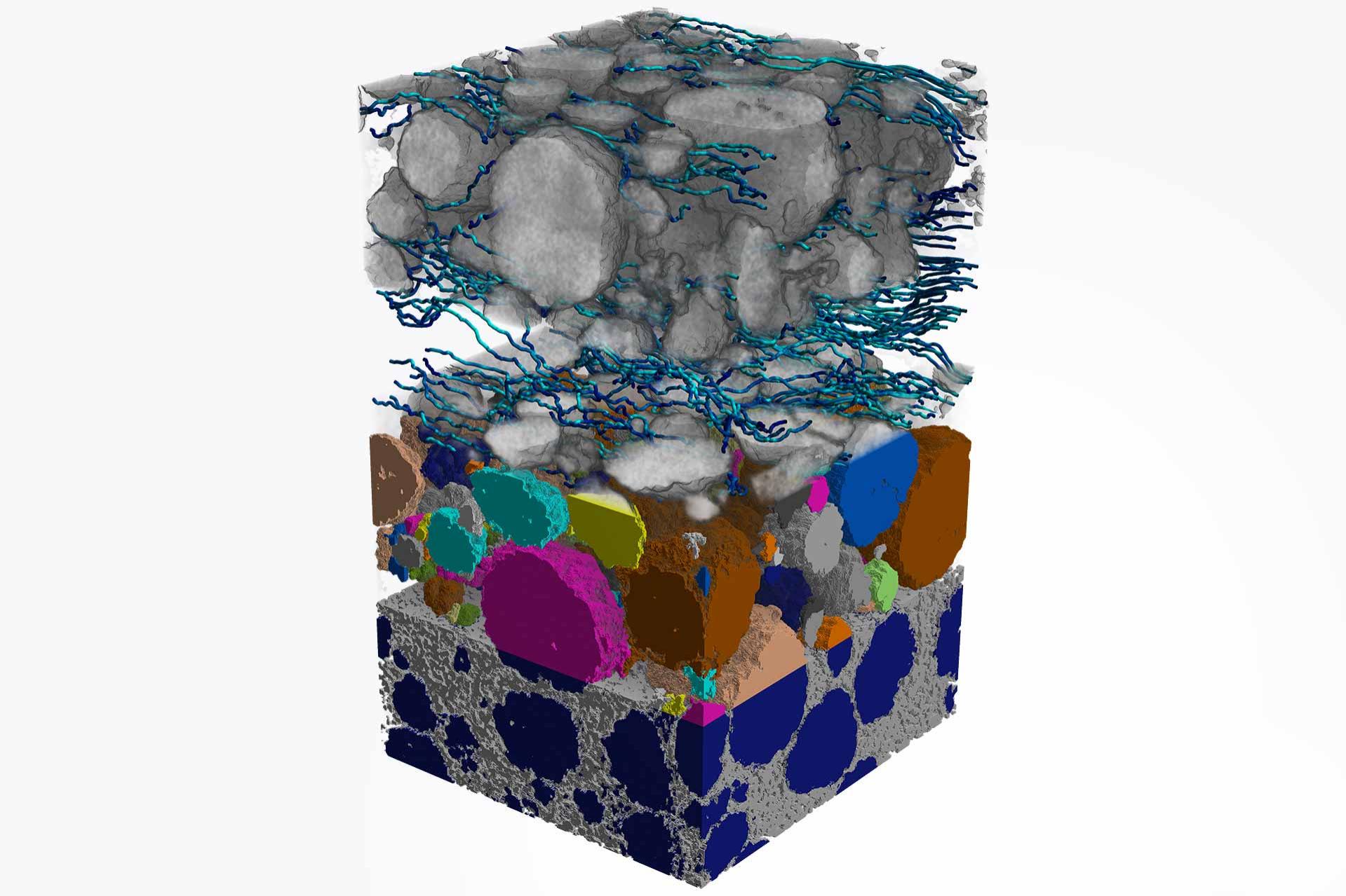


Two types of AI-driven image segmentation
 segmentation of battery layers")
 segmentation of battery layers")
Semantic (Pixel-based) segmentation of battery layers, using an AI-driven model in ZEISS arivis Cloud
 segmentation of granular structure within a battery layer")
 segmentation of granular structure within a battery layer")
Instance (object based) segmentation of granular structure within a battery layer, using an AI-driven model in ZEISS arivis Cloud


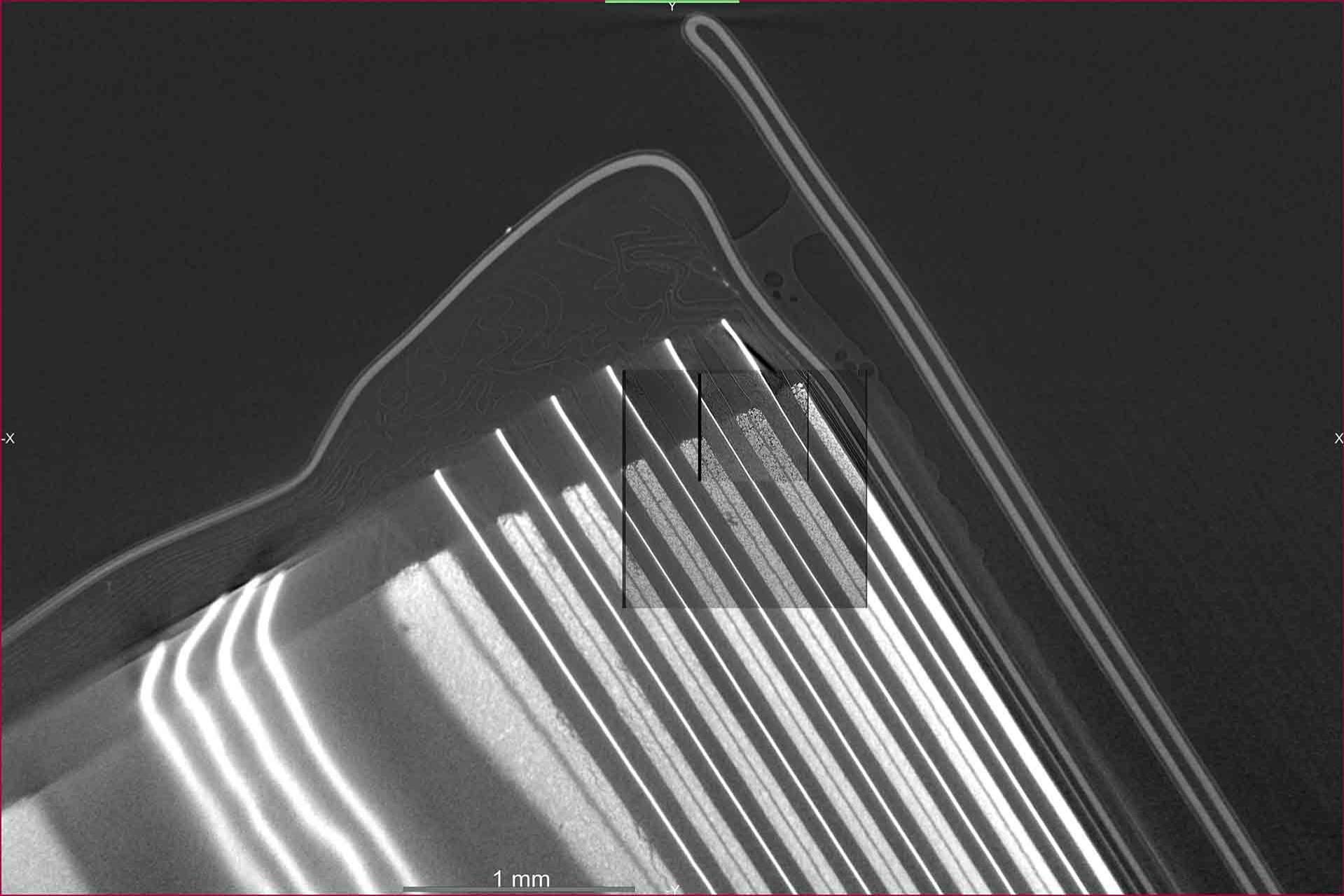
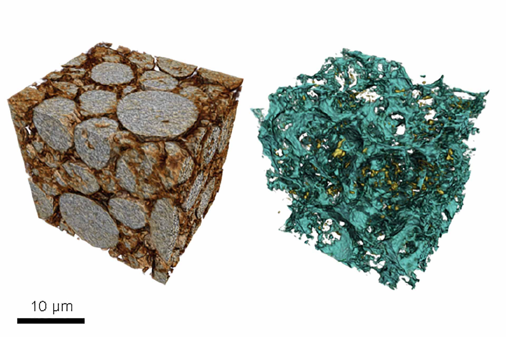
The Inlens EsB signal (right) compared to the Inlens SE signal (left) provides added material contrast between graphite and silicon and reveals the ceramic coating on both sides of the polymer separator.


