ZEISS Elyra 7
Product

ZEISS Elyra 7​

Live Imaging with Unprecedented Resolution down to Molecular Details​

ZEISS Elyra 7 includes a wealth of microscopy techniques to meet your experimental needs across scales, optimally matching resolution, speed, and sensitivity requirements to your demanding samples. Employ SIM Apotome for fast optical sectioning, Lattice SIM for super-resolution imaging, SIM² image reconstruction for resolution excellence down to 60 nm, as well as SMLM and TIRF for investigations at the molecular level. You can combine these techniques to multiply the insights from your specimen and to correlate the acquired data.​

  • Get optical sectioning at exceptional quality.​
  • Resolve structures down to 60 nm.​
  • Explore molecular details with SMLM.​
  • Observe live cell dynamics at up to 255 fps.​
Color-coded projection of Cos-7 cells labeled via immunofluorescence with anti-alpha-Tubulin Alexa 488.

Color-coded projection of Cos-7 cells labeled via immunofluorescence with anti-alpha-Tubulin Alexa 488.

Color-coded projection of Cos-7 cells labeled via immunofluorescence with anti-alpha-Tubulin Alexa 488.

The images demonstrate the excellent sectioning capabilities of SIM² image reconstruction algorithm.

Objective: Plan-Apochromat 100× / 1.57 oil

Resolution Excellence with Lattice SIM²

With SIM², a novel image reconstruction algorithm raising the SIM technology to a new level, you can now double the conventional SIM resolution. Lattice SIM² comes with outstanding out-of-focus light suppression, giving you the sharpest sectioning in widefield microscopy even for highly scattering samples. SIM² image reconstruction robustly reconstructs all structured-illumination-based acquisition data of your Elyra 7 – with minimal artefacts – for living and fixed samples.

Caption: The images demonstrate the excellent sectioning capabilities of SIM² image reconstruction algorithm.

Speed and Efficiency for Your Experiments

While doubling the classic SIM resolution, SIM² gives you gentle imaging of living and fixed specimens at high speeds of up to 255 fps. Combine SIM² with Burst and Leap modes to make super-resolution acquisition faster than ever before. With SIM Apotome mode, even lossless acquisition can be achieved, meaning for every reconstructed image just one raw image is needed! Or make use of Elyra 7 Duolink to image two differently stained structures simultaneously and use the multiple colors to boost resolution even further.

Caption:  Time lapse imaging of the endoplasmic reticulum (Calreticulin-tdTomato) in a Cos-7 cell reveals highly dynamic structural changes.

SMLM: Xenopus laevis A6 cells (epithelial kidney cells).

SMLM: Xenopus laevis A6 cells (epithelial kidney cells).

SMLM: Xenopus laevis A6 cells (epithelial kidney cells).

Gp120, a nuclear pore complex protein arranged with eightfold symmetry was labeled with Alexa Fluor 647.

Flexibility for Your Research

Elyra 7 handles virtually all types of samples, including photo-sensitive cell cultures, scattering C. elegans and plants or tissue sections of up to 100 µm thickness. Elyra 7 includes several microscopy techniques: Lattice SIM², SIM² Apotome, widefield DIC, SMLM and TIRF. You can correlate images of the same sample acquired using any or all of all these techniques to multiply the insights from your specimen. You can even combine Elyra 7 with a variety of other imaging systems such as LSM with Airyscan or scanning electron microscopy in a correlative workflow.

Caption: SMLM: Xenopus laevis A6 cells (epithelial kidney cells).

The Technology Behind It

Lattice SIM: 3D Super-resolution Live Cell Imaging

In Lattice SIM, the sample area is illuminated with a lattice spot pattern instead of grid lines as in conventional SIM. This leads to a dramatic increase in imaging speed. In addition, the lattice pattern provides higher contrast to allow a more robust image reconstruction. Since the sampling efficiency of lattice pattern illumination is 2× higher compared to classic SIM, you need less laser dosage for sample illumination. This lattice illumination makes SIM a preferred live cell imaging technique. The strongly improved photon efficiency of lattice illumination allows you to increase the imaging speed while achieving higher contrast and lower photo dosage.

Images of Cos-7 cell stained with anti-alpha-Tubulin Alexa fluor 488 were processed with the conventional SIM algorithms based on generalized Wiener filter and with the novel SIM² reconstruction. The images show an improvement of resolution for SIM² compared to SIM. Objective: Plan-Apochromat 63× / 1.4 Oil.

Images of Cos-7 cell stained with anti-alpha-Tubulin Alexa fluor 488 were processed with the conventional SIM algorithms based on generalized Wiener filter and with the novel SIM² reconstruction. The images show an improvement of resolution for SIM² compared to SIM. Objective: Plan-Apochromat 63× / 1.4 Oil.

Images of Cos-7 cell stained with anti-alpha-Tubulin Alexa fluor 488 were processed with the conventional SIM algorithms based on generalized Wiener filter and with the novel SIM² reconstruction. The images show an improvement of resolution for SIM² compared to SIM. Objective: Plan-Apochromat 63× / 1.4 Oil.

SIM²: Double Your SIM Resolution

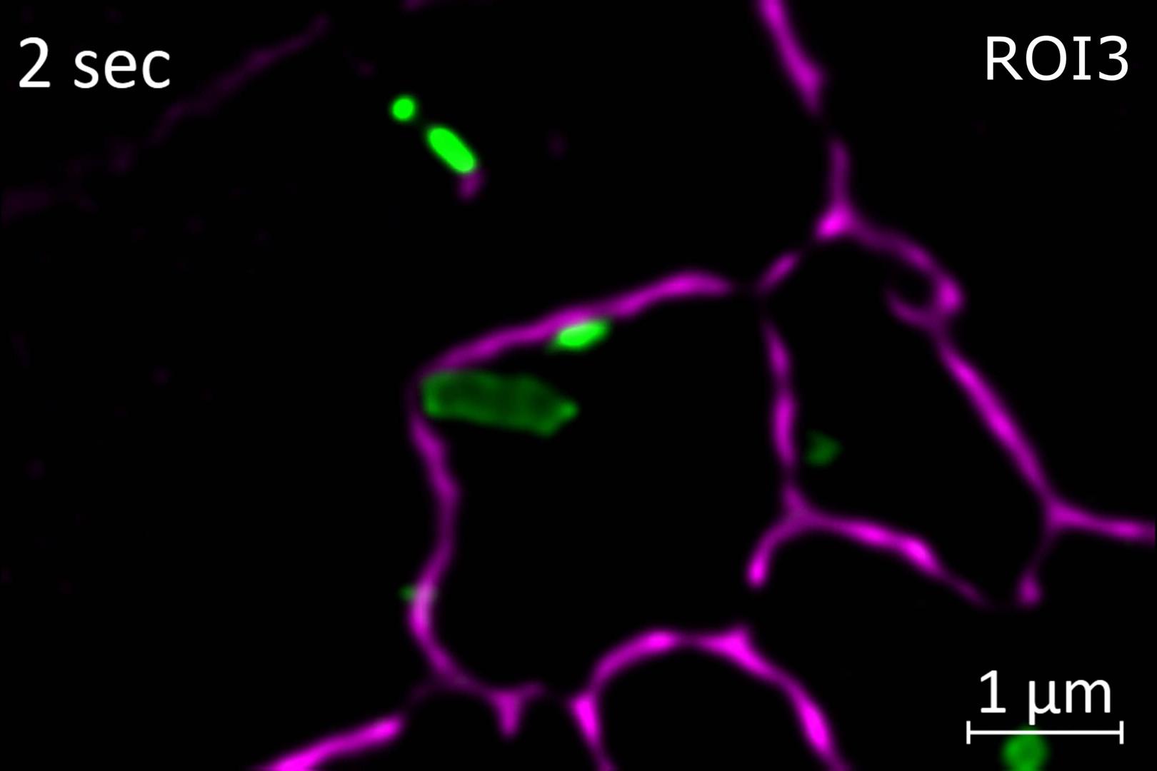
SIM² is the novel, groundbreaking image reconstruction algorithm that increases the resolution and sectioning quality of structured illumination microscopy data. SIM² is compatible with all SIM imaging modes of your Elyra 7 and fully integrated in the ZEN software.

Unlike conventional reconstruction algorithms, SIM² is a two-step image reconstruction algorithm. First, order combination, denoising and frequency suppression filtering are performed. All the effects resulting from these digital image manipulations are translated into a digital SIM point spread function (PSF). The subsequent iterative deconvolution uses this very PSF. Similar to advantages of using experimental PSF for deconvolution of hardware-based microscopy data, the SIM² algorithm is superior to conventional one-step image reconstruction methods in terms of resolution, sectioning and robustness.

Architecture of threefold labeled synaptonemal complexes from mouse testis visualized via immunolabeling of SYCP3 with SeTau647, SYCP1-C with Alexa 488 and SYCP1-N with Alexa 568 and Lattice SIM² mode.
Architecture of threefold labeled synaptonemal complexes from mouse testis visualized via immunolabeling of SYCP3 with SeTau647, SYCP1-C with Alexa 488 and SYCP1-N with Alexa 568 and Lattice SIM² mode. Marie-Christin Spindler, AG Prof Ricardo Benavente, Biocenter of the University of Würzburg.
Marie-Christin Spindler, AG Prof Ricardo Benavente, Biocenter of the University of Würzburg.

Architecture of threefold labeled synaptonemal complexes from mouse testis visualized via immunolabeling of SYCP3 with SeTau647, SYCP1-C with Alexa 488 and SYCP1-N with Alexa 568 and Lattice SIM² mode.

 Marie-Christin Spindler, AG Prof Ricardo Benavente, Biocenter of the University of Würzburg.
Marie-Christin Spindler, AG Prof Ricardo Benavente, Biocenter of the University of Würzburg.

Architecture of threefold labeled synaptonemal complexes from mouse testis visualized via immunolabeling of SYCP3 with SeTau647, SYCP1-C with Alexa 488 and SYCP1-N with Alexa 568 and Lattice SIM² mode.

Multi-color Super-resolution Imaging for Conventionally Stained Samples

Lattice SIM² enables you to perform multi-color imaging at resolution down to 60 nm for conventionally stained samples. Due to its small size, three-color imaging of the synaptonemal complex has previously been possible only using complex methods like super-resolution imaging of three-fold expanded samples. Lattice SIM² resolves the two strands of SYCP3 (lateral elements) as well as SYCP1-C (C-terminus of transverse filaments) without special sample treatment or staining for distances well below 100 nm. More importantly, the three-color image provides structural information about the distances between the proteins SYCP3 and SYCP1. Even within the SYCP1 protein, the different labeled N- and C-Terminus can be clearly separated with less than 50 nm resolution between the two labels.

I remember seeing the first results. I could only laugh as I was simply amazed. My next reaction was to email some of the key users who could immediately benefit. From the tissue neurobiologists, to cell and molecular immunologists, to those working with yeast and those working with bacteria, all of them are already gaining from SIM².

Peter O‘Toole

Head of Imaging and Cytometry, University of York

SIM² Apotome: Comparison of widefield and SIM² Apotome single plane images of Cos-7 cells stained for microtubules (anti-alpha-tubulin Alexa Fluor 488, green) and nuclei (Hoechst, blue). Objective: LD LCI Plan-Apochromat 25× / 0.8 Imm Corr

SIM Apotome: Flexible Optical Sectioning

SIM Apotome
Live cell imaging with a widefield system often suffers from out-of-focus blur or background signal. These effects can decrease contrast and resolution of your images. The SIM Apotome acquisition mode of Elyra 7 uses structured illumination to give you fast optical sectioning with crisp contrast and high lateral and axial resolution.

SIM² Apotome
The SIM Apotome acquisition mode in combination with the SIM² reconstruction algorithm now allows you to further tune the gentleness of your fast live-cell imaging with high contrast and resolution. Or use your new optical sectioning speed to increase your productivity when acquiring large sample areas or large volumes at different magnifications.

Expand Your Possibilities

3D PAINT image of mitochondrial membranes in BSC1 (kidney epithelial cells).

3D PAINT image of mitochondrial membranes in BSC1 (kidney epithelial cells).

3D PAINT image of mitochondrial membranes in BSC1 (kidney epithelial cells).

The outer membrane protein Tomm20 was labeled using Ultivue – I2-650 imaging strand. Reshaped PSF encoding for Z information was used to create a 1.4 µm deep 3D PAINT image.

Objective: alpha Plan-Apochromat 63× / 1.46 Oil

3D PAINT image of mitochondrial membranes in BSC1 (kidney epithelial cells).

Single Molecule Localization Microscopy

3D Imaging at Molecular Resolution

In single-molecule localization microscopy (SMLM), fluorescent molecules are sparsely activated so that only one out of many will be in its on-state within a single point spread function (PSF). This lets you determine its center of mass with a localization precision that far exceeds the extension of the PSF. Once recorded, the molecule is turned to its off-state and the cycle of activation/deactivation is repeated until all molecules are captured. The localizations are plotted in a new image to create the super-resolution image. With Elyra 7 you can use SMLM techniques such as PALM, dSTORM and PAINT to achieve lateral localization precision of 10 – 20 nm. The ZEN software will seamlessly perform the image reconstruction of your data.​

In addition, Elyra 7 provides you with 3D SMLM mode based on PRILM technology. The PSF is reshaped for encoding the Z position so while acquiring only one plane, you get volume information of 1.4 µm depth at 20 – 40 nm axial resolution. Thus, you can acquire 3D data with consistent molecular precision.

  • Elyra 7 Duolink with two Hamamatsu ORCA-Fusion BT cameras
    Elyra 7 Duolink with two Hamamatsu ORCA-Fusion BT cameras

    Elyra 7 Duolink adapter for simultaneous two-color acquisition with two sCMOS cameras (Hamamatsu ORCA-Fusion BT)

    Elyra 7 Duolink adapter for simultaneous two-color acquisition with two sCMOS cameras (Hamamatsu ORCA-Fusion BT)

  • Cos-7 cell expressing the endoplasmic reticulum marker Calreticulin-tdTomato (magenta) and mitochondrial marker Tomm20-mEmerald (green) was simultaneously imaged for two colors. The movie shows high dynamic interactions of the ER and mitochondria.

Elyra 7 Duolink

Simultaneous Two-color Imaging

Investigation of living samples very often focuses on interactions of different proteins or organelles. Simultaneous imaging of the involved structures is key to proper understanding of these highly dynamic processes. Equip your ZEISS Elyra 7 with a Duolink adapter to operate two Hamamatsu ORCA-Fusion sCMOS cameras in parallel and use all the advantages that a widefield-based technology can offer:​

  • ​True simultaneous two-color imaging within your entire field of view – without any delays​
  • Acquisition of a super-resolved real-time snap-shot of an entire living cell by picking a low exposure time​
  • Increased productivity of fixed cell experiments by doubling the information obtained at the same time​
  • Imaging of any possible color combination, with minimal signal crosstalk as enabled by the integrated multi-bandpass emission filters
  • Acquisition of 4-color images without mechanical filter change – making your multi-color experiments even faster​
  • Multi-color SMLM experiments
U2OS cell expressing Rab5-mEmerald (green) and tdTomato tagged Golgi associated transport marker (magenta). Simultaneous dual-color acquisition with an exposure time of 1.5ms / phase for a FOV of 1024 × 1024 pixel (64 µm × 64 µm).

Burst Mode

Super-resolution Imaging at up to 255 fps

The diffusive and especially the ballistic movement of small vesicles in cells can be captured only when super-resolution and high-dynamic imaging are possible at the same time. With the Burst processing of 2D time lapse data, Elyra 7 is able to generate super-resolution images at 255 Hz in a large field of view and even acquire two colors simultaneously in both Lattice SIM and SIM Apotome acquisition modes.

U2OS cell expressing calreticulin-tdTomato to visualize the endoplasmic reticulum. The time series shows a maximum intensity projection of the volume data set.

Leap Mode

Three Times Faster Digital Sectioning

Elyra 7 Leap mode accelerates the volume imaging speed three times and at the same time decreases the light dosage on your sample. While still capturing all the finest details, the entire volume (18 planes) of the U2OS cell expressing Calreticulin-tdTomato was imaged at 38 volumes / min speed in Lattice SIM acquisition mode. For SIM Apotome acquisition mode, you can expect up to three times higher volume imaging speed.

Applications

ZEISS Elyra 7 at Work

  • The Lattice SIM² image of Cos-7 cells labeled with phalloidin Alexa 488 was acquired with a Plan-Apochromat 100× / 1.57 oil objective. Maximum intensity projection of Z stack.
  • Cos-7 cells labeled via immunofluorescence with anti-alpha-Tubulin Alexa 488, shown as color-coded projection.
  • The Lattice SIM² image of Cos-7 cells labeled with phalloidin Alexa 488 was acquired with a Plan-Apochromat 100× / 1.57 oil objective. Maximum intensity projection of Z stack.
    The Lattice SIM² image of Cos-7 cells labeled with phalloidin Alexa 488 was acquired with a Plan-Apochromat 100× / 1.57 oil objective. Maximum intensity projection of Z stack.

    The Lattice SIM² image of Cos-7 cells labeled with phalloidin Alexa 488 was acquired with a Plan-Apochromat 100× / 1.57 oil objective. Maximum intensity projection of Z stack.

    The Lattice SIM² image of Cos-7 cells labeled with phalloidin Alexa 488 was acquired with a Plan-Apochromat 100× / 1.57 oil objective. Maximum intensity projection of Z stack.

  • Cos-7 cells labeled via immunofluorescence with anti-alpha-Tubulin Alexa 488, shown as color-coded projection.
    Cos-7 cells labeled via immunofluorescence with anti-alpha-Tubulin Alexa 488, shown as color-coded projection.

    Cos-7 cells labeled via immunofluorescence with anti-alpha-Tubulin Alexa 488, shown as color-coded projection. The image demonstrates the excellent sectioning capabilities of SIM² image reconstruction algorithm. Objective: Plan-Apochromat 63× / 1.4 oil

    Cos-7 cells labeled via immunofluorescence with anti-alpha-Tubulin Alexa 488, shown as color-coded projection. The image demonstrates the excellent sectioning capabilities of SIM² image reconstruction algorithm. Objective: Plan-Apochromat 63× / 1.4 oil

Studying the Components of the Cytoskeleton

Due to the fine structures of cytoskeleton components, for example the actin network or microtubular filaments, imaging far below 100 nm is often performed with super-resolution techniques. Lattice SIM² allows you to gain much more structural information from your samples compared to conventional SIM techniques. It not only operates with a resolution of down to 60 nm but also provides markedly improved sectioning quality.

  • Simultaneous imaging of the endoplasmic reticulum (Calreticulin-tdTomato, magenta) and microtubules (EMTB-3xGFP, green) in a Cos-7 cell reveals highly dynamic interaction of these organelles. Objective: Plan-Apochromat 63× / 1.4 Oil
  • Living U2OS cell expressing Tomm20-mEmerald were imaged in 3D with a step size of 110 nm. Objective: Plan-Apochromat 63× / 1.4 Oil
  • Simultaneous imaging of the endoplasmic reticulum (Calreticulin-tdTomato, magenta) and microtubules (EMTB-3xGFP, green) in a Cos-7 cell reveals highly dynamic interaction of these organelles. Objective: Plan-Apochromat 63× / 1.4 Oil.
  • U2OS cell expressing Tomm20-mEmerald. Objective: Plan-Apochromat 63× / 1.4 Oil.

Understanding Biological Processes

Due to its unique lattice structural illumination, Elyra 7 combines high speed imaging with incredible light efficiency, low photon dosage and sensitivity. You can observe cellular, subcellular, and even sub-organelle structures in living specimens in 2D and 3D over time. Whether you are interested in the dynamics of mitochondrial movement, fusion and fission or budding of the endoplasmic reticulum, Elyra 7 Lattice SIM² provides you with the necessary live cell compatibility at super-resolution.

  • LLC PK1 cells expressing H2B-mCherry (magenta) and α-Tubulin mEmerald-GFP (green). Data shown as maximum intensity projection of 12 planes over 3.7 µm depth. High sensitivity of the Elyra 7 allows to image the full FOV during the mitosis. Objective: LD LCI Plan-Apochromat 25× / 0.8 Imm Corr
  • Cos-7 cells expressing the endoplasmic reticulum marker Calreticulin-tdTomato. Data shown as maximum intensity projection of 12 planes over 1.4 µm depth. Objective: Plan-Apochromat 40× / 1.4 Oil
  • SIM² Apotome time lapse data of LLC PK1 cells expressing H2B-mCherry (magenta) and α-Tubulin mEmerald-GFP (green). Data shown as maximum intensity projection of 12 planes over 3.7 µm depth. Objective: LD LCI Plan-Apochromat 25× / 0.8 Imm Corr.
  • SIM² Apotome time lapse data of Cos-7 cells expressing the endoplasmic reticulum marker Calreticulin-tdTomato. Data shown as maximum intensity projection of 12 planes over 1.4 µm depth. Objective: Plan-Apochromat 40× / 1.4 Oil.

Excellent Sectioning at Incredible Speed

SIM² Apotome is your flexible live cell imaging method for experiments that do not require the highest spatial resolution but rely instead on excellent sectioning quality. SIM² Apotome is superior to conventional confocal microscopy in terms of lateral and axial resolution as well as volume acquisition speed while it is also very gentle to your sample. The high NA (1.4) 40× magnification images almost reach the resolution and sectioning capabilities of a conventional SIM microscope, while multiplying acquisition speed.

  • Volume tile scan image of a thin mulberry section. Data shown as maximum intensity projection over 11 µm depth. Acquisition time: Below 2 min. Objective: EC Plan-Neofluar 10× / 0.3 air. Sample: “Maulbeere” from TS-Optics Set Dauerpräparate Botanik 25St.
  • Volume tile scan image of a leaf cross section. The image shows a maximum intensity projection of a Z stack. Acquisition time: Below 2 min. Objective: EC Plan-Neofluar 10× / 0.3. Sample: “Leaf” from TS-Optics Set Dauerpräparate Botanik 25St.
  • SIM² Apotome volume tile scan image of a thin mulberry section with an EC Plan-Neofluar 10× / 0.3 air objective. Data shown as maximum intensity projection over 11 µm depth. Sample: “Maulbeere” from TS-Optics Set Dauerpräparate Botanik 25St.
  • SIM² Apotome volume tile scan image of a leaf cross section, imaged with an EC Plan-Neofluar 10× / 0.3 objective. The image shows a maximum intensity projection of a Z stack. Sample: “Leaf” from TS-Optics Set Dauerpräparate Botanik 25St.

Fast Tile Scanning of Very Large Areas

The high-speed performance of SIM Apotome acquisition mode allows for fast tile scan imaging of very large areas at excellent sectioning quality. A mulberry section of 11.1 mm² × 11 µm size was imaged using Nyquist sampling in all three directions and in two colors in less than 2 minutes. Similar speeds also were achieved for a cross section of a leaf.

  • Cos-7 cell expressing EMTB-3xGFP (green) and EB3-tdTomato (magenta) shows dynamic movement of microtubules. Imaged in Lattice SIM 9 phase mode. Objective: Plan-Apochromat 63× / 1.4 Oil
  • Actin dynamics in a Cos-7 cell expressing LifeAct-tdTomato were imaged with the SIM Apotome 3D Leap mode over time. The image shows a maximum intensity projection of 30 planes over 3.4 µm depth. Objective: Plan-Apochromat 40× / 1.4 Oil
  • Cos-7 cell expressing EMTB-3xGFP (green) and EB3-tdTomato (magenta) shows dynamic movement of microtubules. Imaged in Lattice SIM 9 phase mode.
  • Actin dynamics in a Cos-7 cell expressing LifeAct-tdTomato were imaged with the SIM Apotome 3D Leap mode over time.

Defining Specific Needs for Speed and Resolution

The need for higher imaging speeds and decreased light dosage is almost unlimited. The robustness of Elyra 7 structured illumination patterns plus the image reconstruction software allow a significant reduction to the number of phase images required for both Lattice SIM and SIM Apotome acquisition modes, while decreasing the resolution only slightly. Lattice SIM acquisition can be operated at 9 phase images per frame while 3 phase images per frame are sufficient for SIM Apotome, increasing the imaging speed by 44 % and 66 %, respectively.

  • Murine brain expressing the neuronal marker Thy1-eGFP, imaged in Lattice SIM mode over a Z stack range of 75 µm.
  • Zebrafish embryo expressing a vascular marker fli1-EGFP, imaged in SIM Apotome mode over a Z stack range of 100 µm.
  • Murine brain expressing the neuronal marker Thy1-eGFP, imaged in Lattice SIM mode over a Z stack range of 75 µm.
    Murine brain expressing the neuronal marker Thy1-eGFP, imaged in Lattice SIM mode over a Z stack range of 75 µm.  Sample courtesy of Herms Lab (MCN, University of Munich, Germany)
    Sample courtesy of Herms Lab (MCN, University of Munich, Germany)

    Murine brain expressing the neuronal marker Thy1-eGFP, imaged in Lattice SIM mode over a Z stack range of 75 µm.

    Murine brain expressing the neuronal marker Thy1-eGFP, imaged in Lattice SIM mode over a Z stack range of 75 µm.

  • Zebrafish embryo expressing a vascular marker fli1-EGFP, imaged in SIM Apotome mode over a Z stack range of 100 µm.
    Zebrafish embryo expressing a vascular marker fli1-EGFP, imaged in SIM Apotome mode over a Z stack range of 100 µm. Sample courtesy of Haass Lab (MCN, University of Munich, Germany)
    Sample courtesy of Haass Lab (MCN, University of Munich, Germany)

    Zebrafish embryo expressing a vascular marker fli1-EGFP, imaged in SIM Apotome mode over a Z stack range of 100 µm.

    Zebrafish embryo expressing a vascular marker fli1-EGFP, imaged in SIM Apotome mode over a Z stack range of 100 µm.

Resolving Details Hiding in the Depth

Despite being a structured illumination-based microscope, Elyra 7 Lattice SIM² as well as SIM² Apotome also provide you with super-resolution and high-quality sectioning in thick or scattering samples. The combination of robust illumination patterns and excellent image reconstruction technology enabled us to image throughout an entire murine brain section of ~80 µm thickness expressing the neuronal marker Thy1-eGFP.

  • Lattice SIM² 3D image of a C. elegans larvae
  • Living yeast expressing GFP-coupled membrane marker and mCherry-coupled Golgi associated protein shown as maximum intensity projection. Sample courtesy of C. MacDonald, G. Calder & P. O’Toole (Department of Biology & Bioscience Technology Facility, University of York, UK)
  • SIM² Apotome 3D image of a leaf of a living A. thaliana sample shows the microtubules (Tubulin-GFP) in the upper three cell layers. Sample and data curtesy of G. Calder and P. O’Toole (Department of Biology & Bioscience Technology Facility, University of York, UK)
  • Zebrafish embryo expressing a vascular marker fli1-EGFP was imaged in 3D. The figure shows the maximum intensity projection of the tile scan Z stack data set. Objective: Plan-Neofluar 10× / 0.3. Sample courtesy of Haass Lab (MCN, University of Munich, Germany)
  • Lattice SIM² 3D image of a C. elegans larvae. Image shows a maximum intensity projection. Sample courtesy of Mango Lab (University of Basel, Switzerland).
  • Lattice SIM² time lapse images of living yeast expressing GFP-coupled membrane marker and mCherry-coupled Golgi associated protein. Sample courtesy of C. MacDonald, G. Calder & P. O’Toole (Department of Biology & Bioscience Technology Facility, University of York, UK).
  • SIM² Apotome 3D image of a leaf of a living A. thaliana sample shows the microtubules (Tubulin-GFP) in the upper three cell layers. Sample and data curtesy of G. Calder and P. O’Toole (Department of Biology & Bioscience Technology Facility, University of York, UK).
  • SIM² Apotome: Zebrafish embryo expressing a vascular marker fli1-EGFP was imaged in 3D. Maximum intensity projection of the tile scan Z stack data set. Sample courtesy of Haass Lab (MCN, University of Munich, Germany).

Discovering the Diversity of Life

You can investigate living or fixed, small or large, thin or thick specimens using Elyra 7 Lattice SIM², SIM² Apotome or SMLM modes. Whether you study vesicle dynamics in cells or yeast or you want to unravel the architecture of plants, C. elegans, zebrafish, D. melanogaster or bacteria, with Elyra 7 you will experience easily accessible super-resolution imaging for your favorite model organism and many other specimens.

  • SIM² Apotome and Lattice SIM² images of a murine brain expressing the neuronal marker Thy1-eGFP. The images show the color-coded or maximum intensity projections of the volume data.
  • SIM² Apotome and Lattice SIM² images of a murine brain expressing the neuronal marker Thy1-eGFP. The images show the color-coded or maximum intensity projections of the volume data.
  • SIM² Apotome and Lattice SIM² images of a murine brain expressing the neuronal marker Thy1-eGFP. The images show the color-coded or maximum intensity projections of the volume data.
  • SIM² Apotome and Lattice SIM² images of a murine brain expressing the neuronal marker Thy1-eGFP. The images show the color-coded or maximum intensity projections of the volume data.
    SIM² Apotome and Lattice SIM² images of a murine brain expressing the neuronal marker Thy1-eGFP. The images show the color-coded or maximum intensity projections of the volume data. Sample courtesy of Herms Lab (MCN, University of Munich, Germany)
    Sample courtesy of Herms Lab (MCN, University of Munich, Germany)

    SIM² Apotome and Lattice SIM² images of a murine brain expressing the neuronal marker Thy1-eGFP. The images show the color-coded or maximum intensity projections of the volume data.

    Objectives: Plan-Neofluar 10× / 0.3, Plan-Apochromat 40× / 1.4 Oil and Plan-Apochromat 63× / 1.4 Oil.

    SIM² Apotome and Lattice SIM² images of a murine brain expressing the neuronal marker Thy1-eGFP. The images show the color-coded or maximum intensity projections of the volume data.

A Journey Through Different Scales

Biological samples often contain completely different types of information at different length scales. Being able to collect low to high resolution data in the same sample not only makes you more productive but also interconnects the findings and puts them in context to give you the whole picture.

  • SMLM: Eightfold symmetry of the nuclear pore complex in A6 cell.
  • SMLM: Alpha tubulin was labelled with Alexa 555 and beta tubulin with Alexa 488.
  • SMLM: With Elyra 7 you can image a z-depth of 1.4 µm in a single acquisition.
  • SMLM: Eightfold symmetry of the nuclear pore complex in A6 cell.

    SMLM: Eightfold symmetry of the nuclear pore complex in A6 cell.

    SMLM: Eightfold symmetry of the nuclear pore complex in A6 cell.

    Gp210 was labeled with Alexa Fluor 647. Widefield image (1st row, left), SMLM image (1st row, right) and zoomed in region (2nd row, left).

    SMLM: Eightfold symmetry of the nuclear pore complex in A6 cell.

  • SMLM: Alpha tubulin was labelled with Alexa 555 and beta tubulin with Alexa 488.

    SMLM: Alpha tubulin was labelled with Alexa 555 and beta tubulin with Alexa 488.

    SMLM: Alpha tubulin was labelled with Alexa 555 and beta tubulin with Alexa 488.

    The two channels were acquired simultaneously. The epitopes are either occupied by a green fluorophore or by a red one – shown by the mutual exclusion between the green and the red dye molecules.

    SMLM: Alpha tubulin was labelled with Alexa 555 and beta tubulin with Alexa 488.

  • SMLM: With Elyra 7 you can image a z-depth of 1.4 µm in a single acquisition.

    SMLM: With Elyra 7 you can image a z-depth of 1.4 µm in a single acquisition.

    SMLM: With Elyra 7 you can image a z-depth of 1.4 µm in a single acquisition.

    3D SMLM image of Alexa 647 α-tubulin color coded for depth.

    SMLM: With Elyra 7 you can image a z-depth of 1.4 µm in a single acquisition.

Single Molecule Localization Microscopy (SMLM)

SMLM encompasses techniques such as PALM, dSTORM, and PAINT. With high power lasers across the visible spectrum and dual camera detection, Elyra 7 allows researchers to gain access to a broad range of dyes, markers and fluorophores in almost any possible combination.

  • Resolve Molecular Structures -  SMLM allows you to map precise locations of individual proteins.
  • Determine the Relationships Between Molecules -  Detect two channels with molecular precision.
  • Capture Information in Three Dimensions -  Untangle molecular relationships in z with confidence.

Downloads

    • ZEISS Elyra 7 with Lattice SIM²

      Your Live Imaging System with Unprecedented Resolution

      File size: 7 MB
    • ZEISS Lattice SIM Family

      Full Access to Super-Resolution Imaging for all Research Areas

      File size: 3 MB
    • Super-Resolution Imaging by Dual Iterative Structured Illumination Microscopy

      File size: 6 MB
    • ZEISS Elyra 7 and Idylle Everspark Buffer

      Streamlined experiments and reproducible results in localization microscopy

      File size: 1 MB
    • Introducing Lattice SIM for ZEISS Elyra 7

      Structured Illumination Microscopy with a 3D Lattice for Live Cell Imaging

      File size: 1 MB
    • Gamma ZEISS Lattice SIM

      Accesso completo all’imaging a super risoluzione per tutte le aree di ricerca

      File size: 10 MB

Contact ZEISS Microscopy

Contact

Form is loading...

/ 4
Next Step:
  • Step 1
  • Step 2
  • Step 3
Contact us
Required Information
Optional Information

If you want to have more information on data processing at ZEISS please refer to our data privacy notice.