

ZEISS Axio Imager 2 for Materials Research
Open Microscope System for Automated Material AnalysisWhen you perform advanced materials research introduce ease of use into your light microscopy workflow. Benefit from accurate and reproducible results using ZEISS Axio Imager 2 for materials. Choose the system tailored to your applications. Expand your instrument with dedicated solutions for e.g., particle analysis, confocal or correlative microscopy.


Reproducible Results
Enjoy Vibration-Free Working ConditionsStability is essential if you want to obtain good results. Appreciate the stable imaging conditions of Axio Imager 2, especially when working with high magnifications or performing time dependent studies. Due to the motorization of Axio Imager 2 achieve quick and reproducible results while always working under constant conditions.


Modular Design
Gain Enhanced FlexibilityWhether in academic or industrial research, materials microscopy faces various challenges. With Axio Imager 2 you will be able to meet and win these challenges. Attach application-specific components and perform e.g. particle analysis. Investigate non-metallic inclusions (NMI), liquid crystals, or semiconductor-based MEMs. Expand your instrument with dedicated solutions for confocal or correlative microscopy.


High Optical Performance
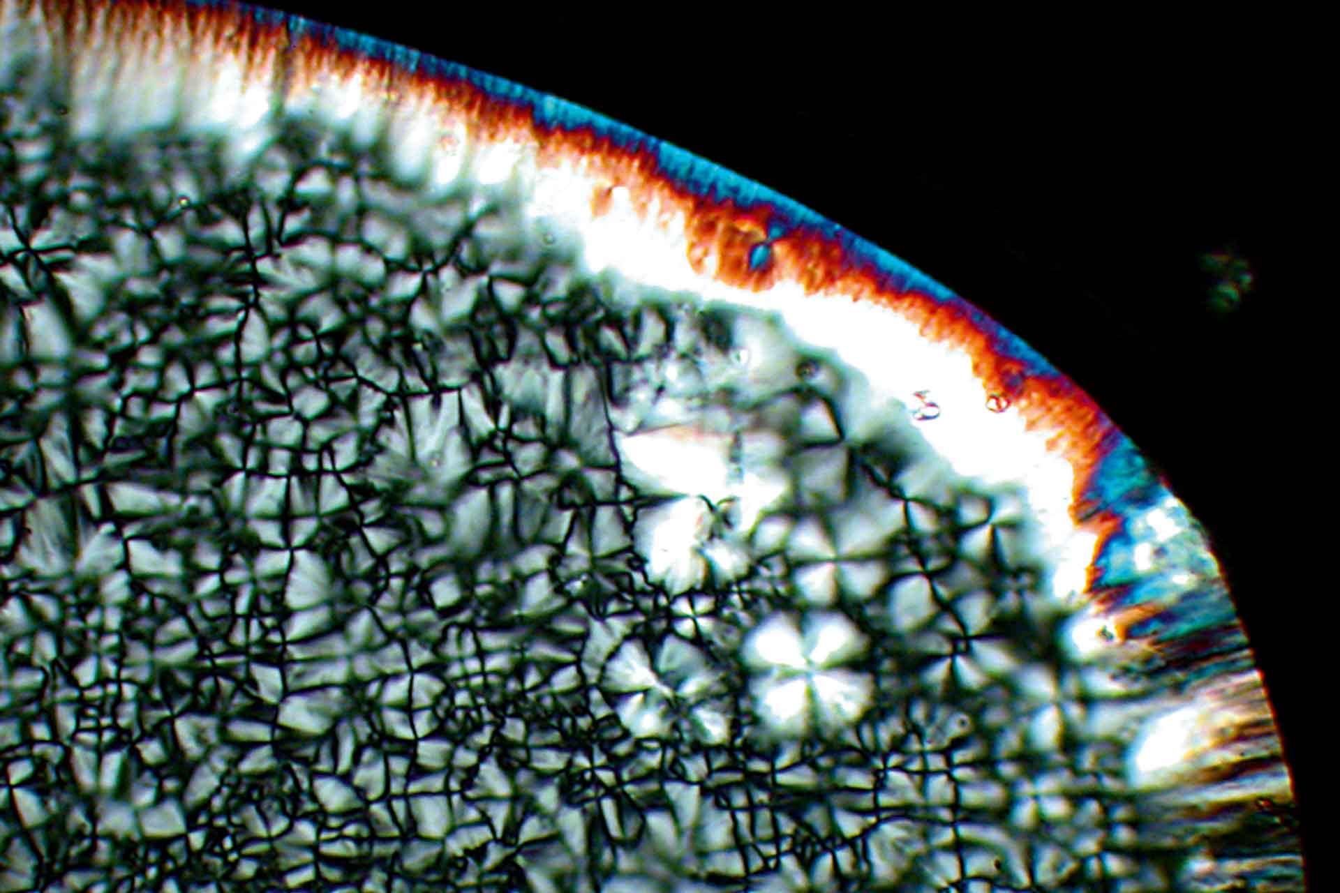
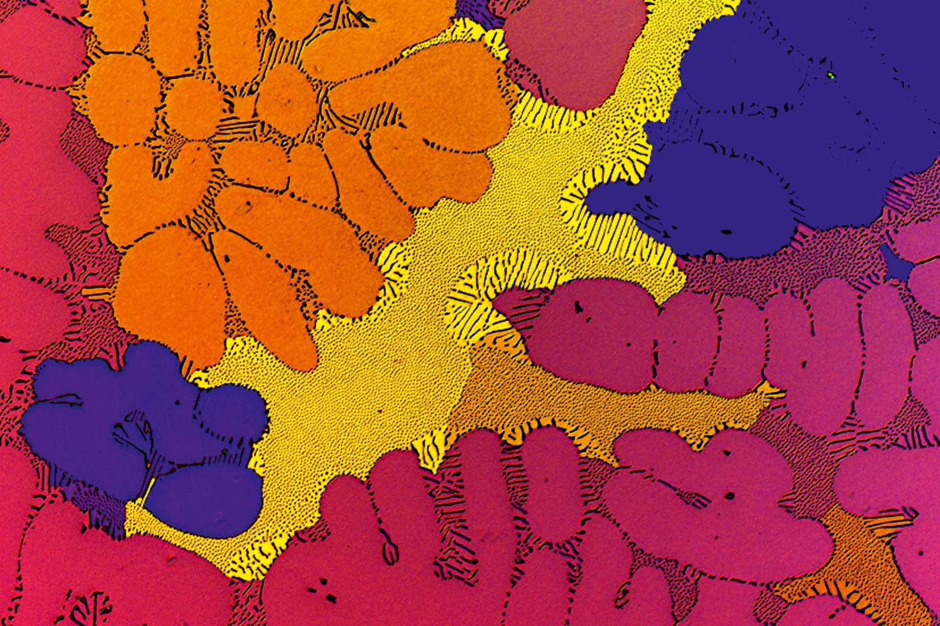
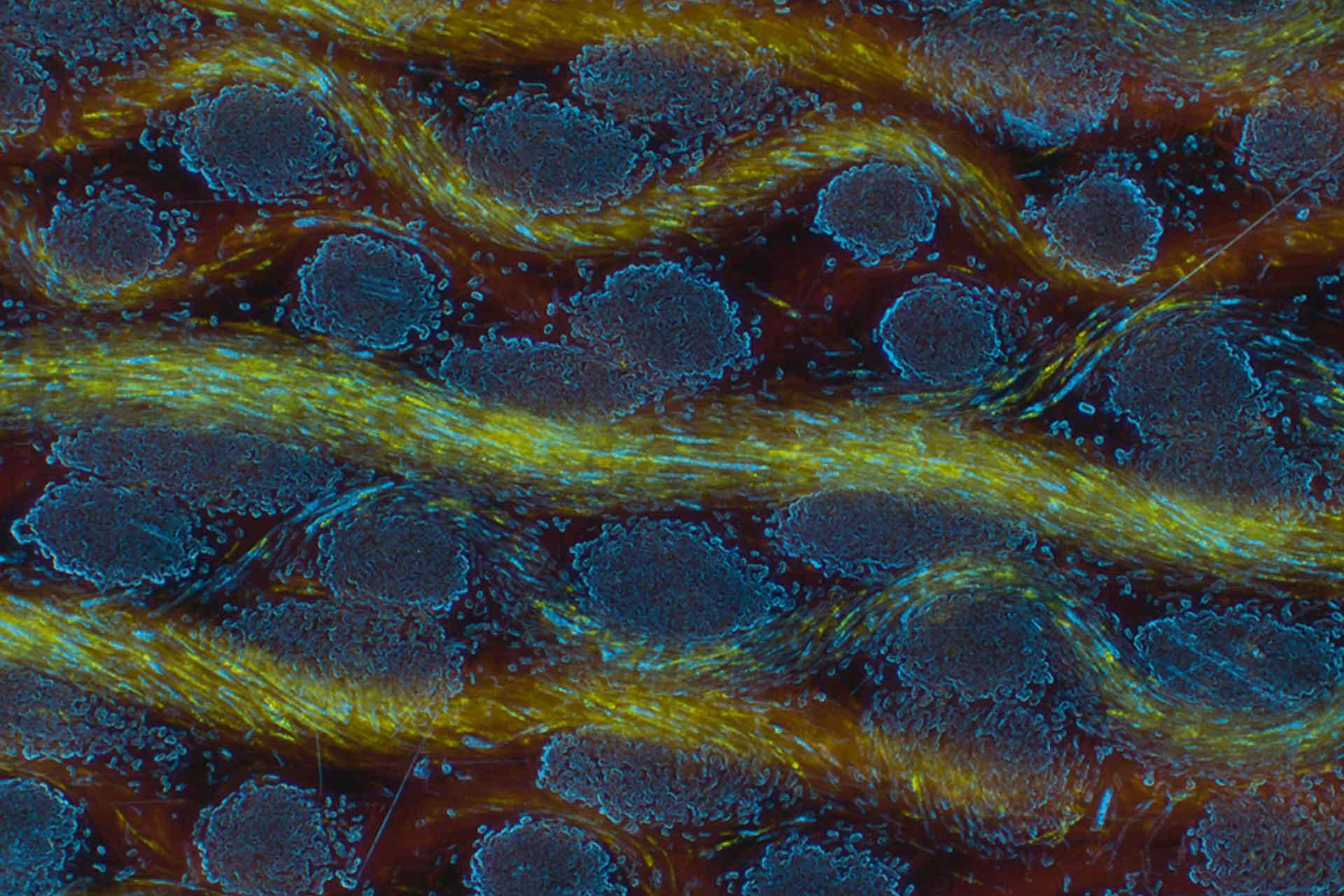
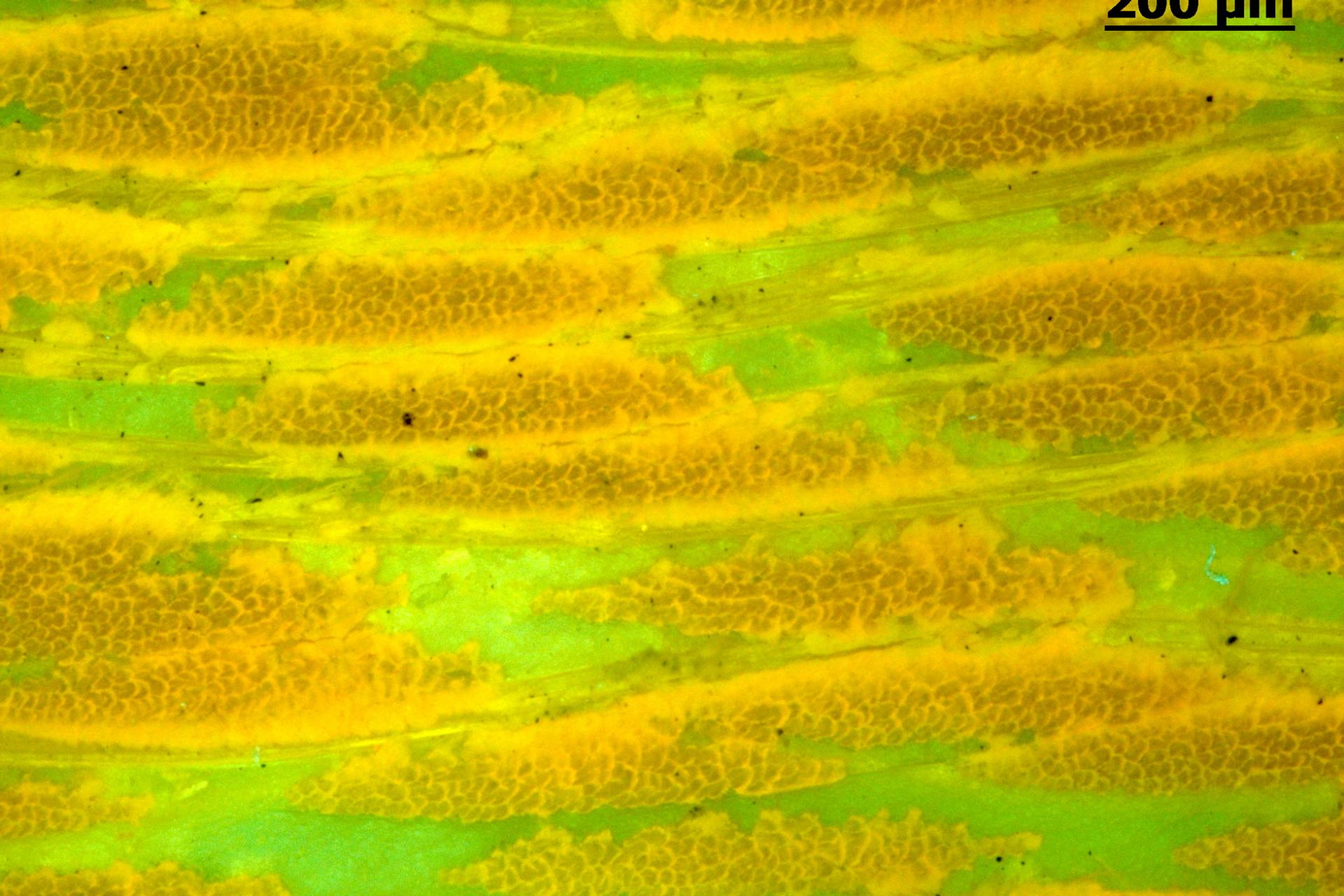
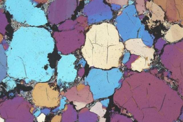
Achieve Superb Contrast and Resolution- Examine a range of materials e.g., metals, composites, or liquid crystals with different contrasting techniques.
- Use reflected light and observe your samples in brightfield, darkfield, differential interference contrast (DIC), circular differential interference contrast (C-DIC), polarization or fluorescence.
- Use transmitted light and examine your samples in brightfield, darkfield, differential interference contrast (DIC), polarization or circular polarization. The contrast manager ensures reproducible illumination settings.


")
")


")
")


")
")


")
")
")
")


Ergonomics
As Multifaceted as Your SurfacesAxio Imager.Z2m or Axio Imager.M2m display key operating functions on a touch screen, giving you fingertip control of all motorized components. Additional control buttons are positioned ergonomically around the focus drive, with tactile surfaces that make them easy to distinguish. Axio Imager.D2m has five pre-programmed buttons while Axio Imager.Z2m has ten user-definable buttons. Axio Imager.A2m is encoded.


Thermomicroscopy
Flexible Documentation of Temperature ChangesDo you want to examine how temperature influences the behavior of metals, crystals, ceramics or plastics?
With ZEN core and Linkam heating stages you define heating or cooling experiments. Document the temperature pattern in a time lapse series. The result you obtain is a log of the temperature and vacuum data in each time lapse image. Observe sample changes during the heating or cooling process for example in the field of quality control.


")
")


