Imaging Applications in Neuroscience Summit

Brought to you by the ZEISS and MPFI Research Partnership

Hybrid event at Max Planck Florida Institute for Neuroscience

Imaging Applications in Neuroscience Summit

From May 16 - 18, 2023, ZEISS and Max Planck Florida Institute (MPFI) brought together neuroscience experts and up-and-coming researchers at our inaugural summit to explore neuroscience imaging applications across light and electron microscopy. In this hybrid event, attendees:  

  • Discovered cutting-edge neuroscience research talks presented by experts in the field
  • Learned about innovative microscopy techniques and sample preparation through methods talks
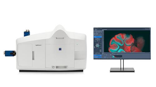
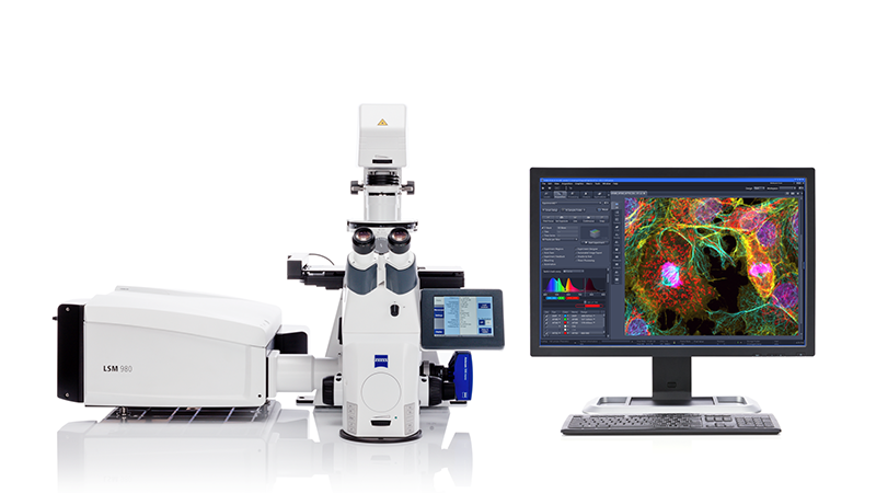
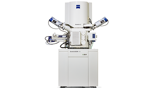
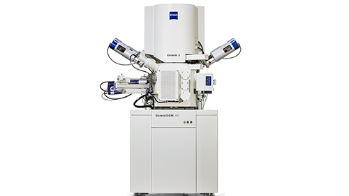
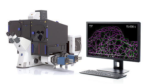
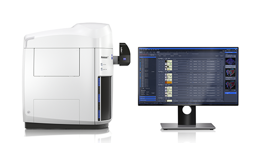
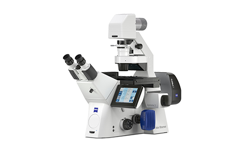
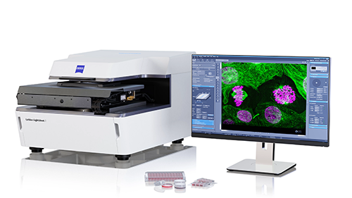
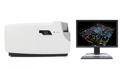
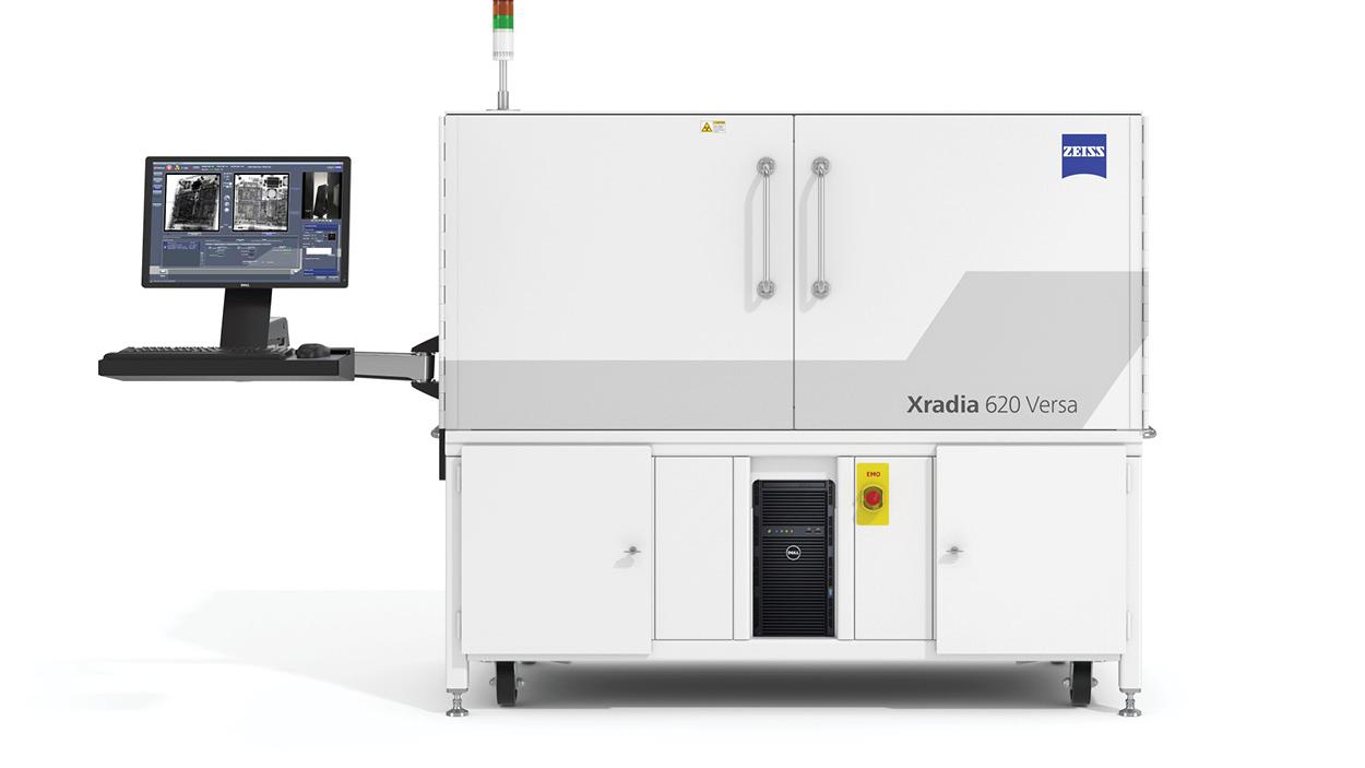
  • Experienced neuroscience imaging applications with state-of-the-art microscope demonstrations
  • Networked with the neuroscience community and spark inspiration in your own research


Learn more about the program and request session recordings below.


Sessions

Browse the topics below to learn more about our sessions. Click on the session to read its abstract.

Access Session Talk Recordings

Please fill out the form below to receive the recordings by email.  
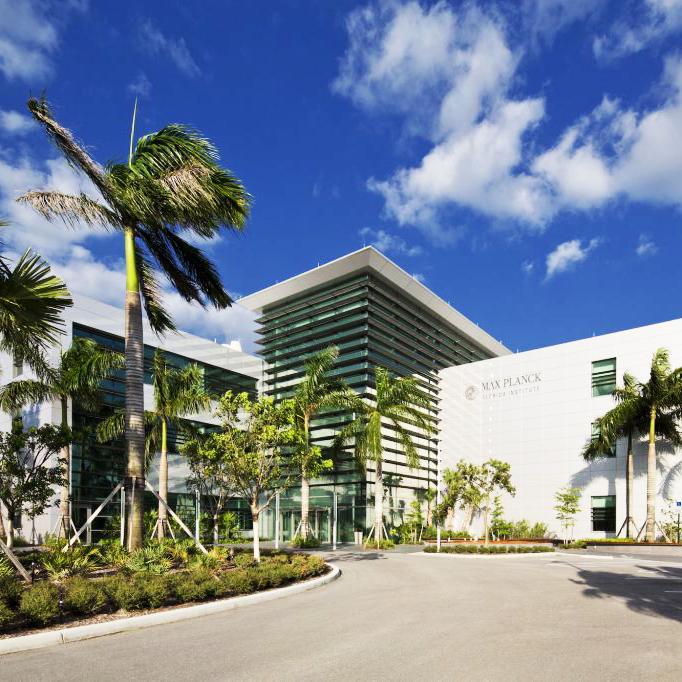
Form is loading...

Imaging Application Demos Showcased at the Summit
  • See the big picture with light sheet imaging of optically cleared whole mouse brains

    Learn how advanced optical clearing techniques and powerful light sheet imaging can accelerate your research. Quickly image a whole cleared mouse brain and visualize brain-wide expression patterns with cellular resolution.

  • Image Science: Progress towards analysis of TB-size images for Neuroscience Applications

    Learn how AI and neuron tracing workflows can leverage your images to quantifiable results, in any modality.

  • Getting more from miniscope calcium imaging: adding multicolor markers to identify neuronal subtypes under the GRIN lens

    Adding cell type markers to your functional GCaMP recordings can add context and enhance interpretations. Here we will demonstrate the pipeline to image up to 10 colors through an implanted GRIN lens on an LSM 980 and co-register these images with miniscope data.

  • Increase your spectral palette and number of markers to gain the most spatial context from your 3D neuroscience applications with the LSM 980

    See how the latest improvements to our LSM 980 spectral capabilities enable the imaging of several markers in the same brain tissue section. New tools such as LSM Plus will further improve resolution, single to noise, and lead to faster acquisition times

  • Introduction to array tomography and serial block face SEM related correlative workflows for neuroscience

    Receive an overview of the correlative light and electron microscopy workflows enabled by Atlas Array Tomography and serial block face scanning electron microscopy. Gain an introduction to the imaging tools and methods used by the technical experts of the MPFI Electron Microscopy Core to explore high resolution ultrastructural and volume imaging in neuroscience samples.

  • Taking a deeper dive into the methods for array tomography and serial block face related correlative workflows

    Join the imaging experts of the MPFI Electron Microscopy Core to take a deeper look the tips, tricks and technical details for preparing and imaging neuroscience samples for array tomography and serial block face SEM related correlative workflows.

  • Go beyond the diffraction limit with ZEISS Elyra 7 with Lattice SIM²

    Explore your neuroscience samples over different scales from high quality overview images to super-resolution of live samples with Lattice SIM², then quantify those results with image analysis tools.

  • Save Time and Money with High Throughput Multiplex Tissue Imaging on the Axioscan 7

    No need to comprimise quality when imaging high quantities. Whole slide multiplex imaging of tissue samples can run 24/7. With an unbiased and flexible filter concept, imaging is not restricted to a proprietary staining kit. Gain further utiliity of your precious samples by keeping it all in context with TIE contrast imaging; revealing the presence and architecture of all cells, stained or unstained. Then seamlessly move to oil-less high magnification imaging for 3D deconvolved data. Axioscan 7 can do it all for you.

  • Automatic Detection, Seamless Tiling and Crisp Optical Sectioning of Multiplexed Mouse Brain on the Axio Observer 7

    Understand global expression and subcellular localization using AI Sample Finder to automatically detect, calibrate and define your ROI. Create seamless whole tissue images followed by high magnification optical sections, while images are all tracked and managed in a single, convenient ZEN Connect project.

  • Discover how accessible lattice light sheet technology really is with ZEISS Lattice Lightsheet 7

    See how to perform rapid volume imaging of even your most sensitive neuroscience samples with next to no phototoxicity and bleaching.

  • One-click Detection, Imaging and Quantification with the Celldiscoverer 7

    With a single click of a button on the system, your multi well plate is detected, calibrated, imaged and quatified. Screening has never been easier.

  • Putting the Synchrotron Beamline in Your Lab with Nondestructive 3D X-ray Microscopy

    Explore 3D tomography insights of whole specimens without dissection or overly complex sample preparation. Gain powerful structural information from your intact biological samples with the high resolution, nondestructive imaging power of ZEISS Xradia Versa 630. See the simple steps to prepare a scan and target your regions of interest for subsequent imaging studies using the easy-to-follow, workflow-guided user interface.

In-Person Location

Max Planck Florida Institute for Neuroscience

Part of the prestigious Max Planck Society based in Germany, MPFI is its first and only institute in North America. Situated in the growing biosciences cluster in scenic Palm Beach County in South Florida, MPFI provides a vibrant, collaborative environment where scientists are provided generous ongoing support to conduct high impact research at the cutting edge.

Attendees had the opportunity to experience the state-of-the-art microscope demonstrations and network with the neuroscience community.