
ZEISS at Cell Bio 2023
Discover the Power of PossibilitiesYour research consumes your thoughts, queuing up questions on how you can see more, see deeper, capture the moment, and make a breakthrough. But what lies beyond your current systems and software capabilities? Discover new innovations to help make the data you seek, visible.
Leverage our experts to help you connect your needs to the right solution, the right training, and the right settings for repeatability.
As your partner, we are focused on sustainability, leveraging our digital efficiencies to fulfill your needs, wherever you are in your consideration process. We are your resource, your network for achieving your next breakthrough. We believe that you are the power behind what's possible, let us help you find innovations to achieve your discoveries, faster.
Join us at ASCB Cell Bio 2023 in booth #515 in bringing together sustainability and cutting-edge technology to display our latest product launch, debut new researcher discoveries on our interactive wall, show our products in a VR showroom, and offer live virtual product introductions.
Tech Talks at Cell Bio
Hear from ZEISS Experts and researchers like you at our 3 Tech Talks. Click on the presentation titles to read the abstracts:-
Exploring Serial Block Face SEM as a Key Technology in the Volume EM Revolution
Presented by Emily Benson, Lead Research Technologist, Cleveland Clinic, and Aubrey Funke, Life Science Electron Microscopy Product Marketing Manager, Carl Zeiss Microscopy
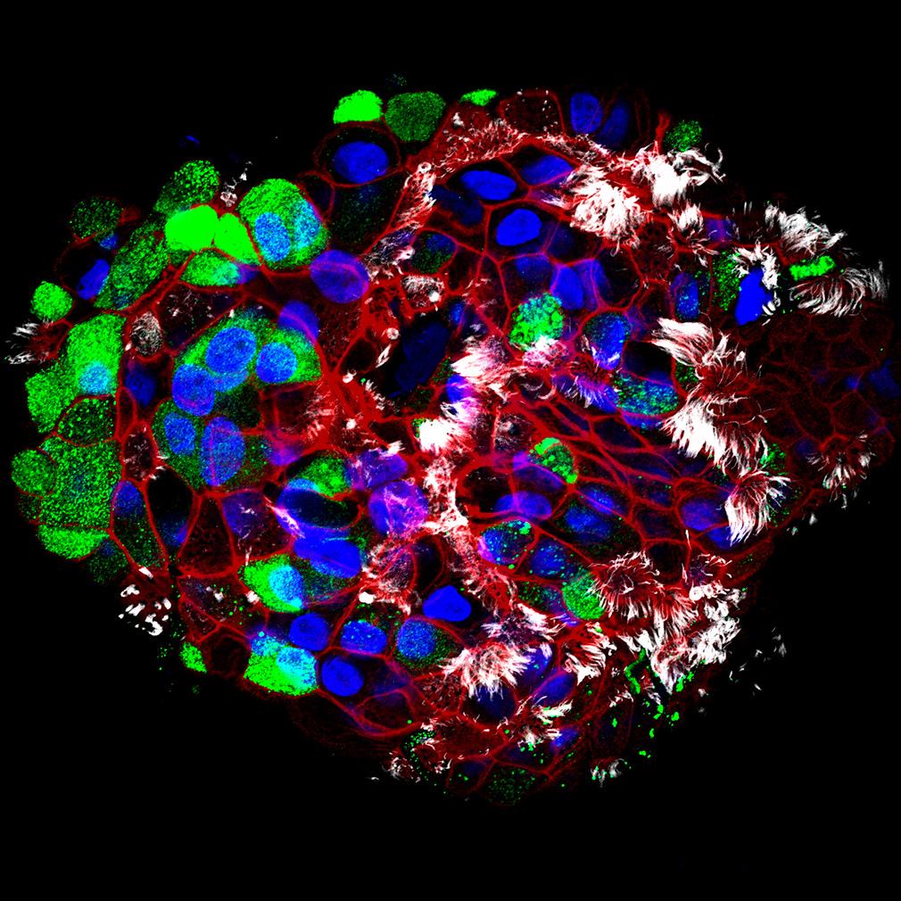
Life takes place in three dimensions. Scanning electron microscopy (SEM) offers opportunities to explore intricate, ultrastructural 3D information of tissues and cells with techniques that are known collectively as volume EM. Join Emily Benson, Lead Research Technologist at Cleveland Clinic, and Aubrey Funke, Life Science Electron Microscopy Product Marketing Manager at Carl Zeiss Microscopy, to discover why volume EM has been named a key technology to watch in 2023. Learn about the ultrastructural imaging power of these individual techniques and how volume EM can spark inspiration in your own cell biology research. Receive an introduction to the growing Volume EM Community and discover how Emily Benson uses serial block face imaging as a key technology in her electron microscopy core throughout a variety of life science applications. Discover the latest volume EM solution from ZEISS. ZEISS Volutome is the next generation of serial block-face imaging: highly automated, fully integrated and optimized for volume data creation. See the features that save you time while allowing you to access large volume data with superb image quality, at low kV.
-
New microscopy tools to profile the dynamics of the gap junction nexus and other molecular mechanisms of brain connectivity
Presented by Randy Stout, Ph.D., Associate Professor, Department of Biomedical Sciences, New York Institute of Technology
Connexin proteins connect cells in all major human organs. The most widely studied connexin, Cx43, is important in diseases of heart, bone, and brain. Cx43 has a short half-life (~3hrs) and interesting but complicated cellular trafficking, membrane localization, channel opening/closing regulation, and endocytosis characteristics. Along with other labs, we have used techniques such as Fluorescence Recovery After Photobleach (FRAP) and others to study the dynamics of gap junction supramolecular protein complexes and the Cx43 protein binding partners that make up the Gap Junction Nexus. We found that the structure and dynamics of gap junctions depends on the connexin type and can transition between stability states depending on cell physiology. Evidence from numerous labs indicates spatial organization within the gap junction nexus that is of size scales and protein densities that limit utility standard approaches in this area of research. We recently turned to the new Dynamics Profiler tool to gain insights into gap junction cell biology that are not possible with other techniques. This has allowed us to obtain data measuring protein concentration, mobility, and diffusion characteristics in smaller spaces and a wider range of labeling densities. These data made possible with Dynamics Profiler, along with other techniques, will be critical to better understanding intercellular communication in tissues, and for building a useful computational model of human cells.
-
Introducing the newest family of ZEISS Imaging Systems for Super-resolution across scales
Presented by Kirstin Elgass, Ph.D., Product Marketing Manager, Carl Zeiss Microscopy
The ZEISS Lattice SIM super-resolution technology has taken research beyond the diffraction limit of conventional microscopy, giving you gentle super-resolution imaging with incredibly high speed and the ability to image deeper into challenging samples beyond 120 nm. Lattice SIM2 uses a 2D Lattice pattern that only requires translation, no rotation, for improved speed and higher contrast for deeper penetration into samples. SIM Apotome mode uses 2D striped illumination also without rotation for ultra-fast, exceptional optical sectioning. In combination with our SIM² reconstruction algorithm, both technologies keep pushing SIM to a new level.
Technology that enables imaging the details of biological samples with super-resolution is great, but each application comes with specific challenges and requirements, for example: imaging speed to capture dynamics, large fields of view and excellent 3D capabilities for contextual imaging of larger samples, sensitivity to image delicate samples, or resolution at the edge of what's physically possible to reveal subcellular ultrastructure.
If you wish you could meet these requirements for each of your samples in the best possible way, while remaining flexible across scales, then join us at ASCB to be among the first to be introduced to a new family of ZEISS imaging systems that will allow you to reveal cellular behavior and inter-cellular dynamics, the vibrant sub-organelle network of life, and life across scales-down to molecular details.
Discover what's new at ZEISS
Attend a workflow session to learn more about our latest microscopy solutions:

Introducing the newest family of ZEISS Imaging Systems for Super-resolution across scales
Be among the first to be introduced to a new family of ZEISS imaging systems that will allow you to reveal cellular behavior and inter-cellular dynamics, the vibrant sub-organelle network of life, and life across scales-down to molecular details. Request a session by filling out the form below.
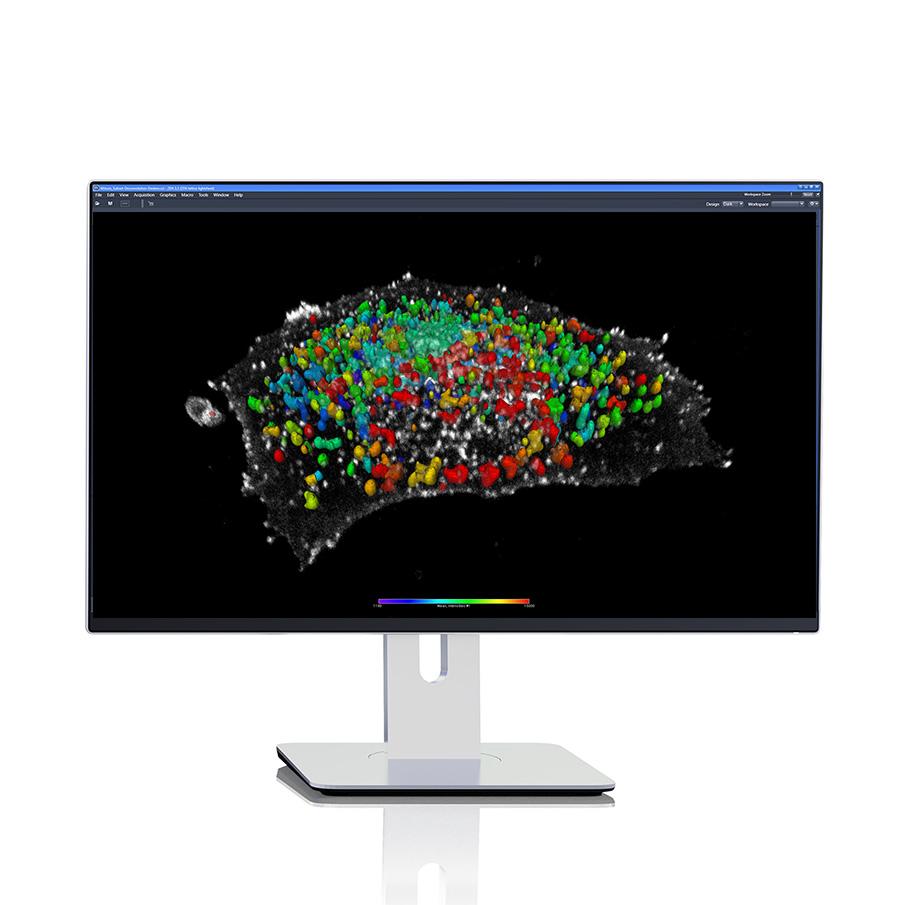
Discover new molecular insights from your live samples with the new Dynamics Profiler
Uncover molecular concentration, asymmetric diffusion, and flow dynamics of fluorescent proteins in your living samples in a single, easy measurement. Develop a more in-depth profile of the molecules in your current experiments, from cell cultures to organoids to whole organisms. Learn how to make these measurements with ease using the new Dynamics Profiler for Airyscan. Request a session by filling out the form below.
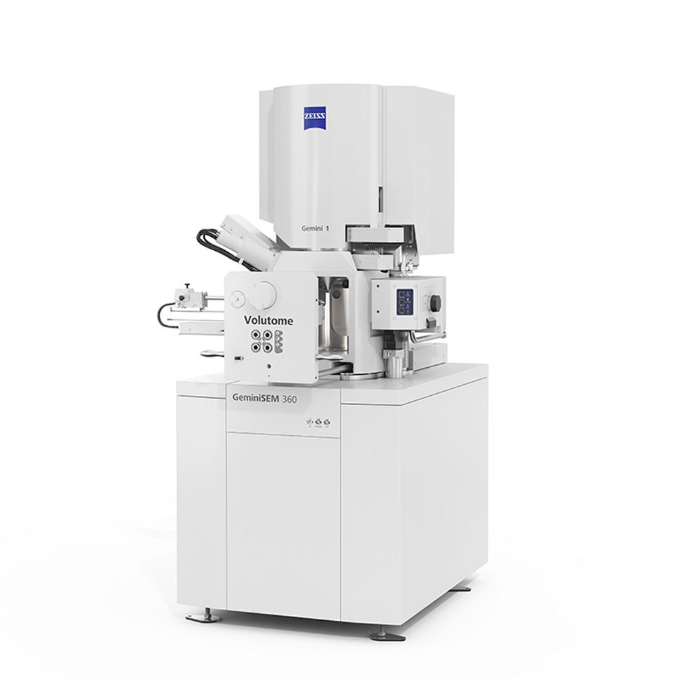

Exploring the ultrastructural imaging power of serial block face SEM
Understand cellular ultrastructure in the 3D context with ZEISS Volutome, your next generation in-chamber ultramicrotome for serial block-face SEM. See how the fully integrated hardware to software stage solution automates cutting, image acquisition and image pre-processing to save you time. From image acquisition to 3D results, experience the latest volume EM solution from ZEISS. Request a session by filling out the form below.

Experience the potential of image analysis solutions, from traditional methods to tailored AI
Learn new ways to improve your work and achieve better results using the ZEISS arivis portfolio of software. Walk through our building blocks for analysis including established operators, custom code, open-source AI models, and customized deep learning models – all while handling data up to terabytes in size. Explore collaboration tools, VR options and scalable automated analysis. Drop by booth #515 anytime for a consultation with one of our image data scientists.