


ZEISS Microscopy Applications Hub
Discover solutions tailored to your researchExplore user stories, application notes, and more!
Skip to main content

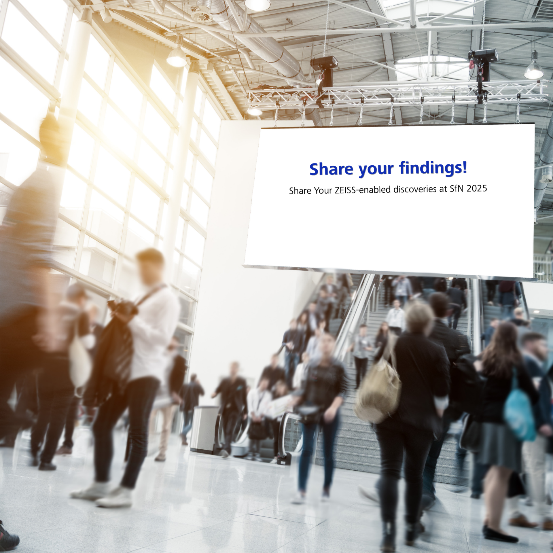
The precision and complexity of neuroscience demand more than just good tools — they demand the right partners. At ZEISS, we’re dedicated to supporting your research journey, from imaging and analysis to training and collaboration. This year in San Diego, explore how ZEISS is shaping the future of neuroscience.
What awaits you at Booth #1213:
We can't wait to see you in San Diego!
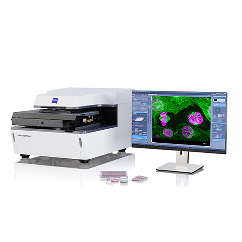
Join us for a focused session exploring the next generation of microscopy. The Lightfield 4D Microsymposium brings together developers, innovators, and early adopters to discuss how this technology is advancing true 3D volumetric imaging at biological timescales - and what this enables in neuroscience.
Stop by Booth #1213 on Monday, November 17 @ 2:30 pm to join the conversation!
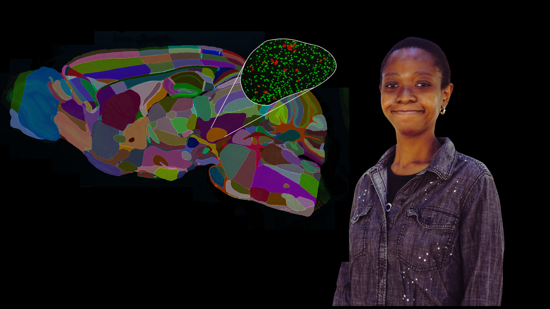
Leading researchers take the stage to share their latest findings — then, together with ZEISS specialists, they dive into the imaging and analysis approaches that made these discoveries possible. Each session pairs scientific excellence with technical expertise, concluding with a live demonstration showing how these workflows can be applied in your own research.
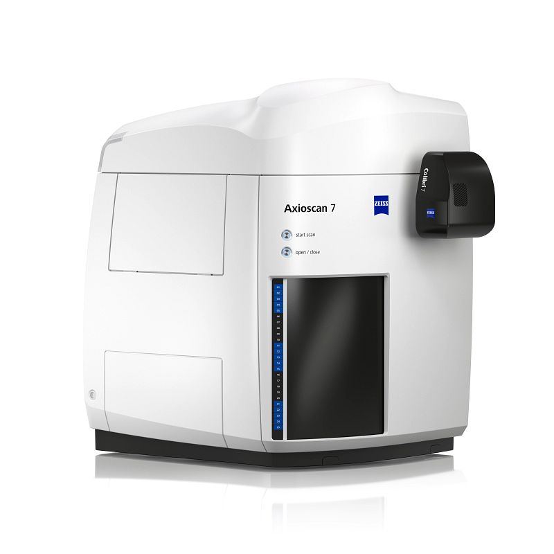
Advance your research with expert support from ZEISS. From high-content imaging to atlas registration, spine detection, and glial morphology analysis, our microscopy and analysis specialists help you get from data acquisition to insights faster.
Capture dynamic biological processes in 3D — instantly. See how Lightfield 4D delivers fast, volumetric imaging for living samples and complex systems, revealing structure and activity in a single acquisition.


ZEISS is evolving beyond instruments — building a connected learning experience for every stage of your research.