
The Voices of X-ray Microscopists
From students to post-docs, to professors and research scientists, X-ray microscopy is transforming the way people see and understand 3D morphology at the microscopic scale.
Enjoy the videos below extracted from our recent programs 3D in 3 Minutes and TomographyTuesday. Learn how scientists in diverse fields are using XRM to make innovative new discoveries.
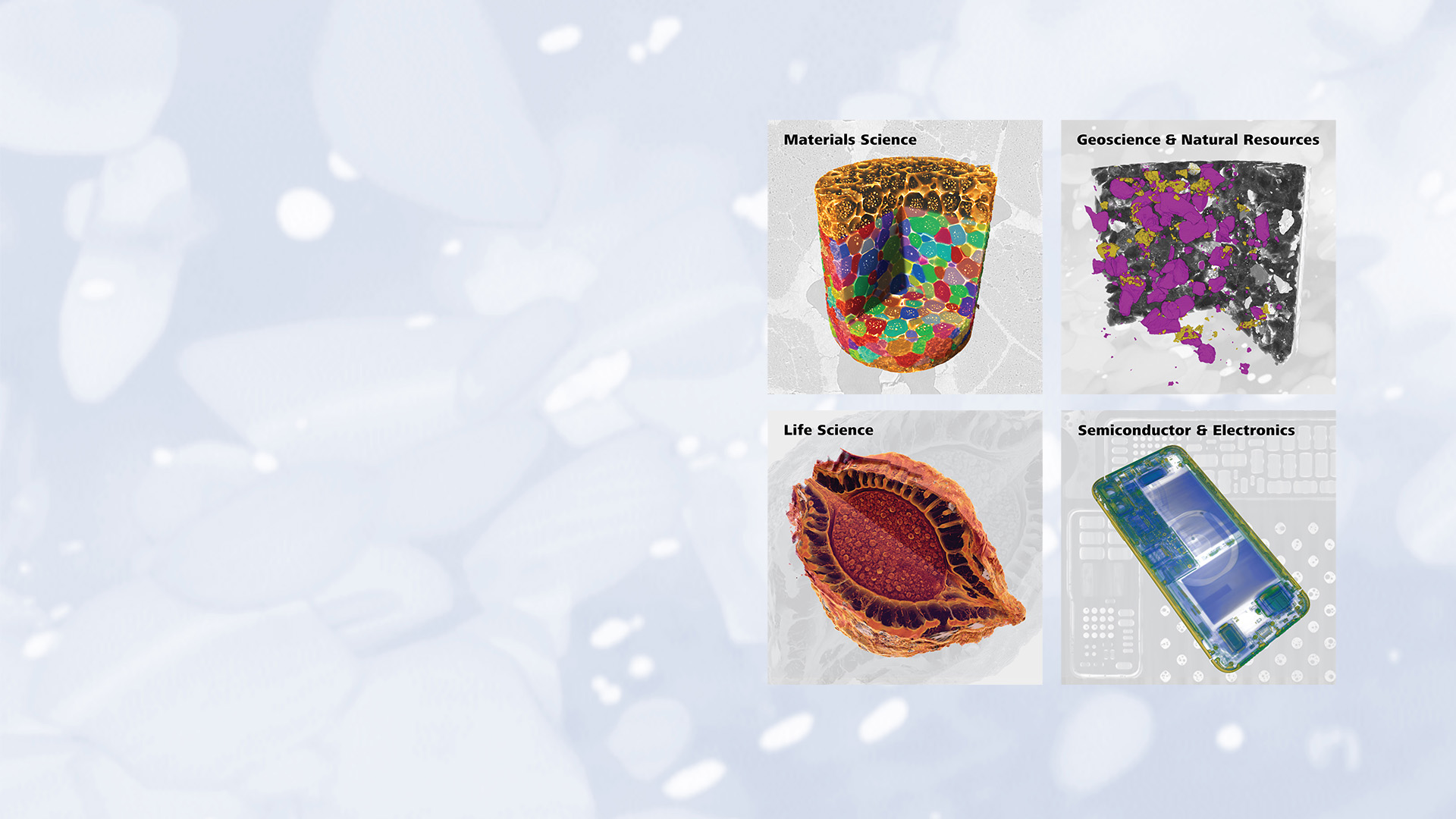
Browse the selection of researchers and topics below, or select an application to jump straight to examples of XRM being used in your field of study.
Enabling the Research Community with Nancy Muyanja

Dr. Nancy Muyanja
XRM & EM Specialist
University of Michigan
Dr. Muyanja is developing the University of Michigan’s X-ray microscopy user base, educating and surprising them with how XRM can solve problems in ways they didn’t know were possible. #3Din3Minutes.
Launching a Materials Research Career with Sridhar Niverty


Dr. Sridhar Niverty
Post Doctoral Research Associate
Purdue University
Dr. Sridhar Niverty showcases some of the diverse 3D imaging projects associated with his PhD and Post-Doc under Prof. Nik Chawla at Purdue University. #3Din3Minutes.
Running an XRM Lab with Yara & Nicholas


Yara Suleiman & Nicolas May
PhD Candidates
University of Connecticut
Yara and Nick handle a diverse range of projects in the University of Connecticut’s REFINE lab under Prof. Sina Shahbazmohamadi. They discuss the key role of XRM in their lab. #3Din3Minutes.
Analyzing Metals Microstructure with Ashwin Shahani


Ashwin Shahani
Associate Professor
University of Michigan
Prof. Shahani from the University of Michigan shares his work, published in Acta Materialia, on studying grain boundary and triple junction networks in polycrystalline materials. #TomographyTuesday.
Watching Cracks in Porous AM Metal with Philip Noell

Philip Noell
Principal Member, Technical Staff
Sandia National Lab
Sandia National Lab, with the University of Utah, are using XRM to study how the deformation and cracking of metal AM parts is influenced by porosity #TomographyTuesday.
Linking Structure to Properties with Dane Wedgeworth

Dane Wedgeworth
US Army Engineer Research
& Development Center
The distribution and structure of the multiple phases in a composite material are critical to performance. See how XRM can help visualize and quantify these key attributes. #TomographyTuesday.
Measuring Fiber Composites with Facundo Sosa-Rey

Facundo Sosa-Rey
PhD Candidate
Polytechnique Montreal
Fiber length and orientation play key roles in the mechanical properties of fiber-reinforced composites. See how the team at Polytechnique Montreal characterized individual fibers in additively manufactured composites. #TomographyTuesday.
See Glass at the Nanoscale with John McCloy & Team

Natalie Smith-Gray, John McCloy, John Bussey
McCloy Research Group
Washington State University
Prof. McCloy’s team from Washington State University shows how nanoscale XRM can be used to characterize structure and degradation in nuclear glass and refractory materials. #TomographyTuesday.
Tracking Transport in Porous Media with Laura Dalton

Laura Dalton
Associate Professor
Duke University
Prof. Dalton shows how '4D' XRM can add an element of time to observe the progression of complex transport phenomena in porous materials. #TomographyTuesday.
Watching Batteries in Real-Time with Paul Choi


Paul Choi
PhD Candidate
Carnegie Mellon University
Paul conducts research in energy materials in Prof. Litster’s lab at CMU. They use nanoscale X-ray imaging, with pioneering in operando setups, to address some of the tough microstructural challenges in electrochemical devices. #3Din3Minutes.
Taking New Views on Plant Science with Keith Duncan


Keith Duncan
Research Scientist
Donald Danforth Plant Science Center
In this video, Keith shares a snippet of the societal significance of plant science, demonstrating how XRM can provide a better understanding of floral development, and therefore yield, in critical crops. #3Din3Minutes.
Understanding Nature’s Mechanics with Gianluca Tozzi

Dr. Gianluca Tozzi
Reader
University of Portsmouth
Dr. Tozzi focuses on the critical need for X-ray microscopy in bioengineering research, including applications of phase contrast imaging and ‘4D’ studies with digital volume correlation. #3Din3Minutes.
Deriving Inspiration from Nature with Ria Mitchell


Dr. Ria Mitchell
Experimental Officer
University of Sheffield
Dr. Ria Mitchell shows how XRM can be used to support bio-inspired engineering, by observing the natural world to understand how to mimic similar structures and functions in man-made materials. #3Din3Minutes.