
Conquer the Confocal
A ZMCC SkillBuilder WorkshopWorkshop Overview & Agenda
This workshop will cover methods and supporting theory to enable attendees to become more comfortable and competent operating their ZEISS LSM 900s and 980s. Prior LSM imaging background will be helpful but not required. It is recommended that attendees secure time on their instruments after the workshop sessions to apply the shared methods on their own challenging samples. Attendees are strongly encouraged to share their images from individual sessions and interact with our Applications team through independent email threads for further optimization.
We will host this workshop at the ZEISS Microscopy Customer Center (ZMCC) in Dublin, CA. This is a two-and-a-half-day in-person paid workshop consisting of presentations to highlight fundamental microscopy concepts as well as hands-on instrument time with trainers to click through your workflows step by step. With small group sizes of <5 attendees/trainer, you will get valuable, individualized access to the most cutting-edge microscopy solutions ZEISS offers.
Topics that will be covered in this workshop include:
Introduction to Confocal
- What is a pinhole and how does optical sectioning work
- Why pixel size matters
- LSM 900 and 980 Lightpaths
- Varying Imaging Conditions
- Dynamic Range
- Speed v. Spectral Separation
- Sampling and averaging
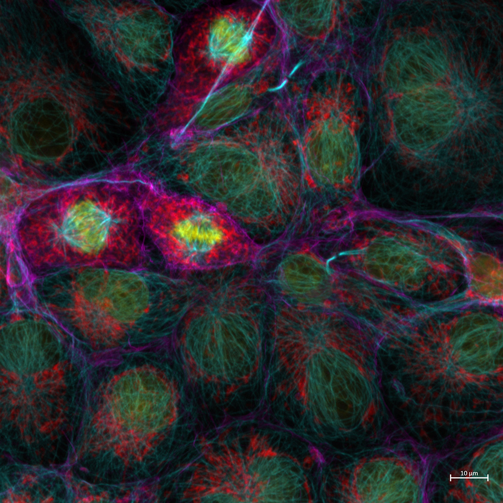
Confocal Imaging Applications
- Z-stacks and axial sampling
- Tiles, positions, and focus strategies
- Time lapse imaging
- Photo manipulation module (FRET/FRAP applications)
Spectral Imaging
- LSM 980 beam path and detector overview (PMTs; Spectral GaAsP; NIR detectors)
- Far red sensitivity on NIR
- Linear Unmixing
- Spectral imaging in Lambda Mode
- Online Fingerprinting
Airyscan Imaging
- Basics of the Airyscan detector
- AS detector alignment
- Superresolution and multiplex modes
- Multiplex modes on LSM 980 (4Y and 8Y)
- Imaging with live cells on the Airyscan
Who should attend?
- Existing users of ZEISS LSMs (700/800/880/900/980)
- Infrequent LSM users looking for a refresher on basic and advanced LSM techniques
- Lab managers or technicians looking to improve their own skill level to better support and/or train other users of the instrument
Learning objectives after completion of this workshop, attendees should:
- Possess an improved ability to select imaging parameters to yield high-quality images of their samples
- Have greater confidence with complex imaging setups, optimizing imaging parameters, troubleshooting on LSMs and analyzing acquired datasets
- Be connected with the North American Applications team who can support them as they work towards acquiring the best data possible
Workshop Package and Payment:
- This is a paid training.
- The investment for this 2.5-day workshop is $3,000. You will receive a link to complete payment by credit card after signing up. If you require a PO to purchase training please reach out to us after registering.
- This package includes:
- World-class training and certification at the end of workshop
- Hotel accommodation within walking distance
- Lunches and refreshments during the workshop
- To guarantee your spot and hotel coverage, please make sure to submit your payment by March 12. Attendees are responsible for arranging transportation to the workshop location.
- Once we have received payment, we will send you a confirmation e-mail detailing hotel information, workshop itinerary, and arrival instructions.
Register for the workshop here.

Dr. Duffy spent the beginning of her career training as a tissue engineer and applying advanced 3D imaging techniques to quantify engineered organs-on-a-chip before becoming an applications specialist for ZEISS. During this time, she established key cellular attachment, proliferation, and migration assays used to assess in-house developed bioinks, and she used a suite of ZEISS microscopes to assess cellularization of 3D printed scaffolds (Celldiscoverer 7, LSM 880, LSM 980, Lightsheet 7). She joined ZEISS in 2020 as the Lattice Lightsheet 7 specialist, and has been a part of the Applications team at the Microscopy Customer Center since 2022 where she spends her time specializing on LSMs and live cell applications.
She earned her B.S. in Biomedical Engineering from VCU in 2011, and completed her PhD in Biomedical Engineering at Carnegie Mellon University in 2016.

Joining ZEISS more than 40 years ago as a Scientific Programmer, Elise began her career programming our Light and Electron Microscopes. She holds a BS in both Chemistry and Mathematics from Binghamton University and obtained her master’s in computer science from Pace University. After 3 years of programming, she transitioned into the Product Management group where she spent the next 10 years as the Product Manager for Image Analysis. She then joined the field Sales team as the Field Product Specialist for Confocal Microscopy at ZEISS, in the Mid-Atlantic Region, before becoming the Head of the NA Product Application Sales Specialist group.