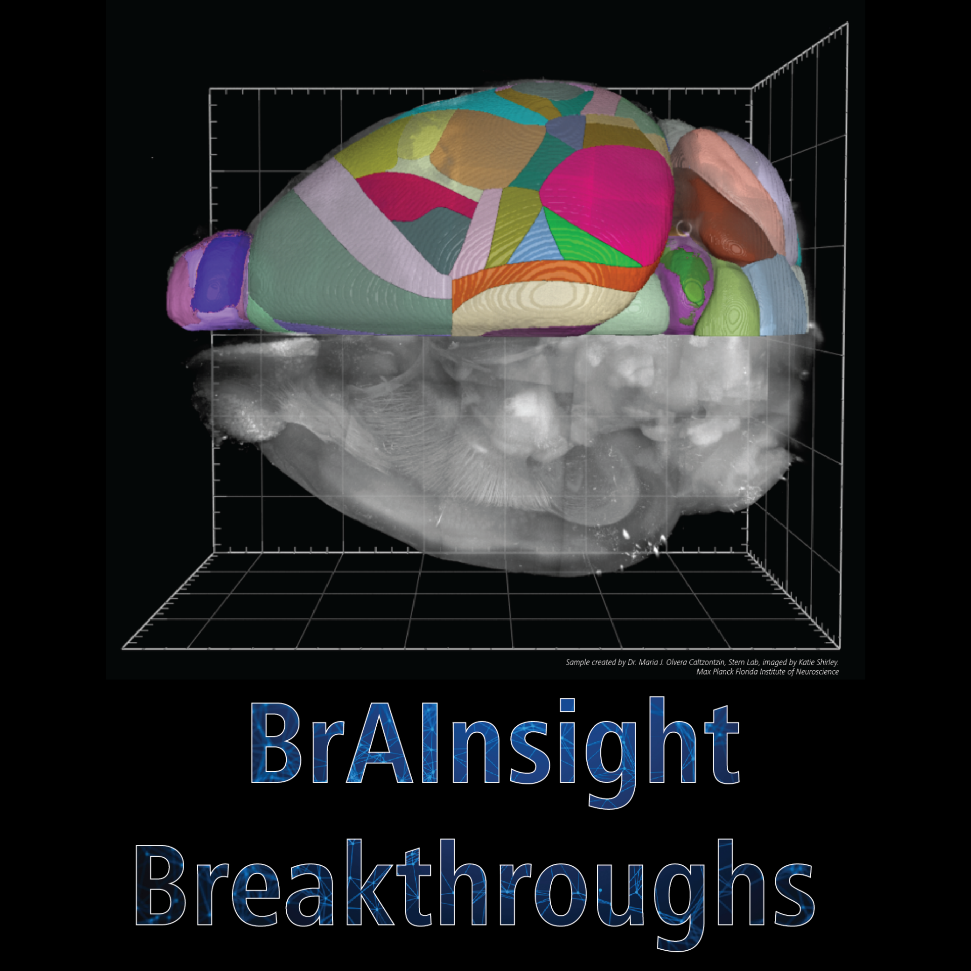

The World is Not Flat - Leveraging 3D whole brain imaging and analysis to investigate the pathophysiology of murine Traumatic Brain Injury
Dr. Mehwish Anwer- 00 years
- 00 months
- 00 days
- 00 hours
- 00 minutes
- 00 seconds
BrAInsight Breakthroughs Webinar Series
Upcoming Webinar Information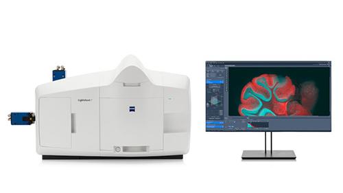


The World is Not Flat - Leveraging 3D whole brain imaging and analysis to investigate the pathophysiology of murine Traumatic Brain Injury
Join us on March 27th for the first webinar in our BrAInsight Breakthroughs Webinar Series. Don't miss this opportunity to learn from neuroscience researchers like you and learn how ZEISS can help revolutionize your work.
Abstract:
Traumatic brain injury (TBI) induces a myriad of pathological processes resulting in brain-wide damage to cellular activity, axonal connectivity, and vasculature integrity. Despite the fundamental importance of understanding impaired brain activity exhibited in post-traumatic epilepsy and other neurological impairments associated with TBI, knowledge of how brain injury affects neuronal activity remains remarkably incomplete. We developed a whole-brain imaging and analysis approach to identify alterations in neuronal activity after TBI as a complementary method to conventional two-dimensional histological approaches. We established an easy-to-follow experimental pipeline to quantify changes in the whole mouse brain using tissue clearing, light sheet microscopy and an optimised open-access atlas registration workflow.
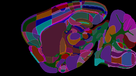
Using the CHIMERA (Closed-Head Impact Model of Engineered Rotational Acceleration) TBI model, TRAP2 mice were subjected to repeated mild TBI or sham treatment followed by tamoxifen injection to lock c-Fos activity after TBI. Brains were SHIELD fixed and passively cleared for imaging of c-Fos+ cells throughout the rostro-caudal axis of the brain using a light sheet microscope equipped with a specialized whole-brain imaging chamber. Volumetric images were stitched and 3D rendered using Zeiss arivis Pro image analysis software. For quantitative analysis, 2D image stacks were exported to segment c-Fos+ cells and register them to the Allen Mouse Brain Atlas using the BrainQuant3D python package. As a result, c-Fos+ cell counts were estimated throughout the brain and heatmaps were generated. A brain-wide reduction in c-Fos cell density was identified in the TBI group compared to sham controls, indicative of TBI-induced changes in whole brain neuronal activity. Further studies using multi-dimensional imaging of axonal and vascular damage coupled with 3D analysis tools will deepen our understanding of post-TBI brain-wide dynamics.
Read more about this topic in a preprint here.


Dr. Mehwish Anwer is a Postdoctoral Fellow in the Department of Pathology and Laboratory Medicine, University of British Columbia, Vancouver, Canada. Dr. Anwer is carrying out her research in Dr Cheryl Wellington’s team at the Djavad Mowafaghian Centre for Brain Health. Before moving to Canada, Dr Anwer carried out her doctoral studies at the University of Eastern Finland for which she received the prestigious European union funded Marie Skłodowska Curie PhD Scholarship. Dr Anwer is investigating the aftermath of Traumatic brain injury (TBI), which is one of the most difficult acquired neurological conditions to treat, due to heterogeneity in nature of impact and evolution of pathology and is identified as a risk factor for development of dementias such as Alzheimer’s disease. To understand the underlying TBI-induced complex pathology, Dr Anwer employs cutting-edge techniques such as tissue clearing, light sheet imaging and spatial transcriptomics. Dr Anwer serves on various UBC committees to advocate integration of principles of equity, diversity and inclusion, and she believes that mutual respect and kindness can go a long way!
Educational Resources
Following the webinar, interact online with resources that will help you to recreate the workflows demonstrated! These resources include protocols and instructional videos regarding:
- Clear brain tissue
- Image whole brains using the Lightsheet 7
- Identify cells of interest
- Register your brain, and cell counts, to the Allen Brain Atlas