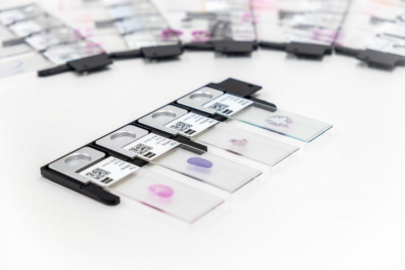

Biopharma Insights Hub
Discover Tailored ApplicationsImaging Innovations for Drug Discovery
See more, discover faster: advance your discovery with our excellence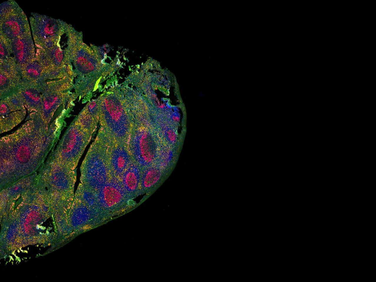


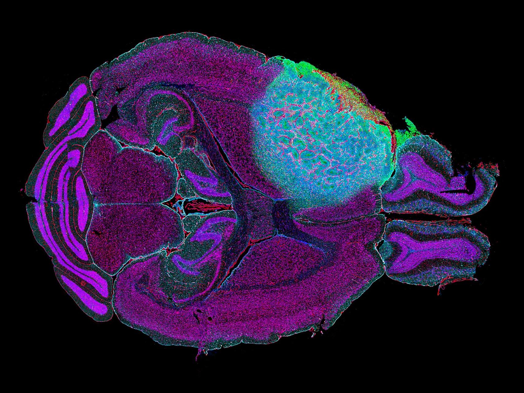
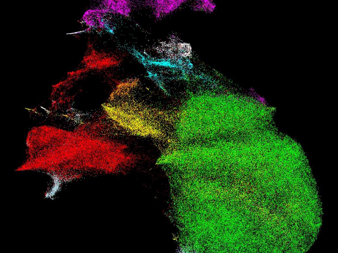
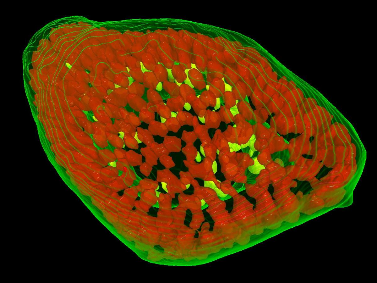
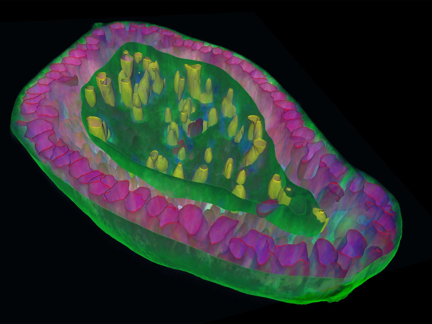
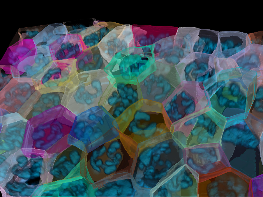
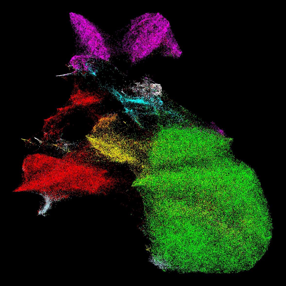
3D Multiplexing Spatial Omics Workflows in Neuroscience
This case study provides a blueprint for the analysis of 3D spatial omics multiplexing datasets from a CODEX multiplex antibody panel imaged with a custom set-up using the Akoya Phenocycler and ZEISS LSM 980 confocal microscope to explore the expression of an extensive list of pre-synaptic markers in the mouse brain.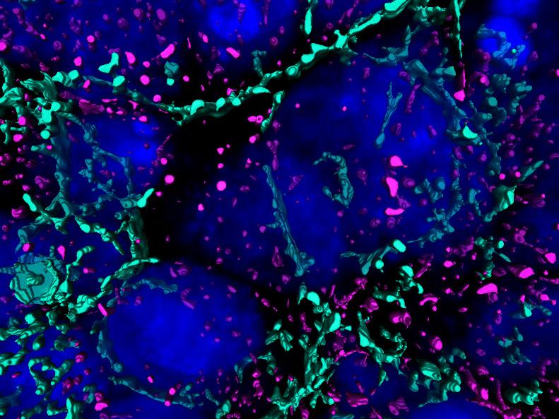

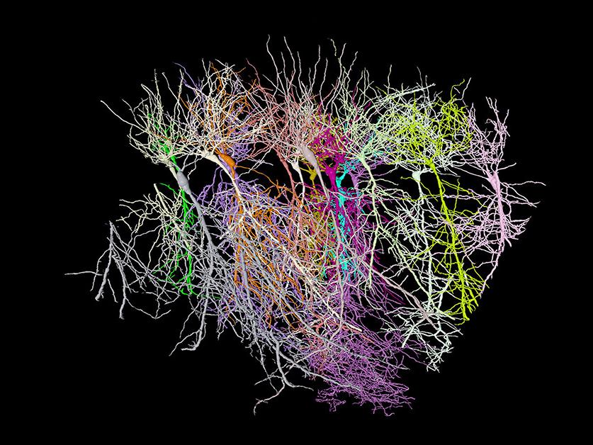

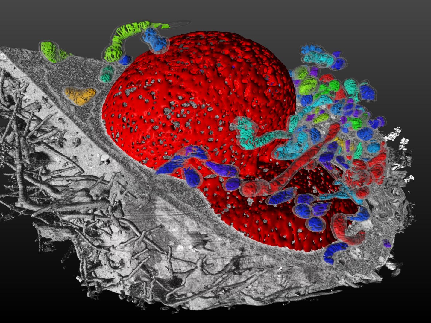
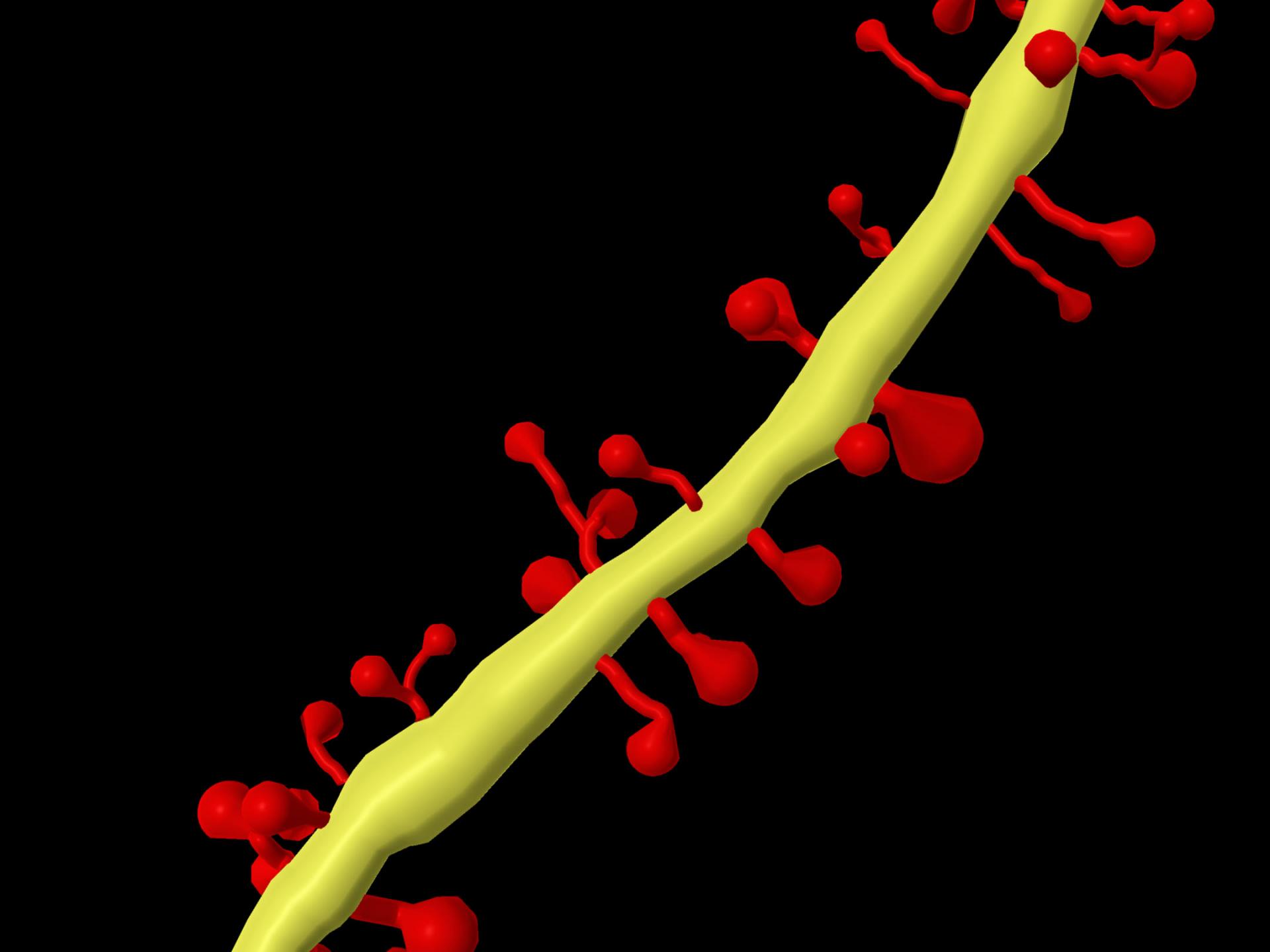
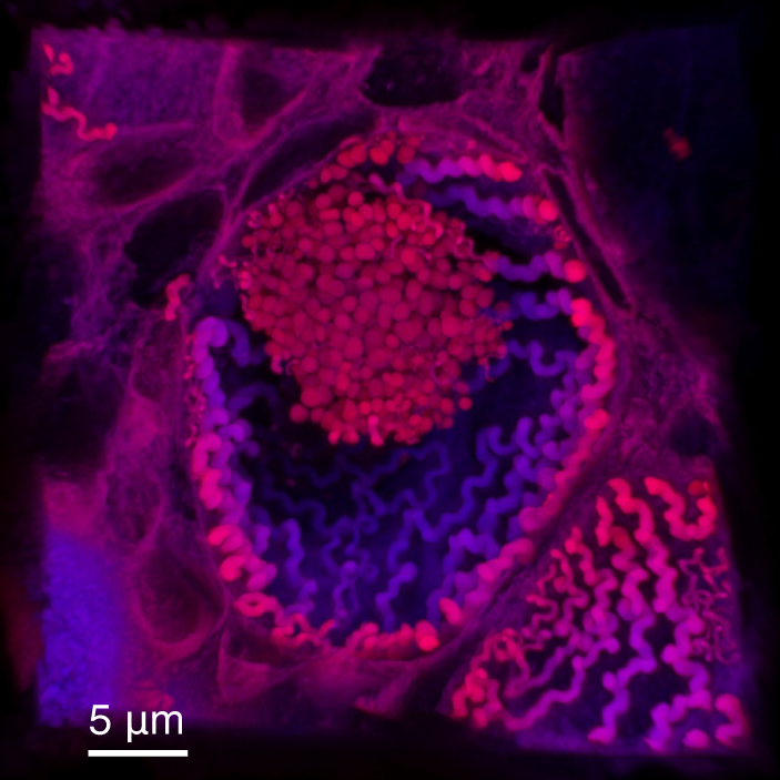
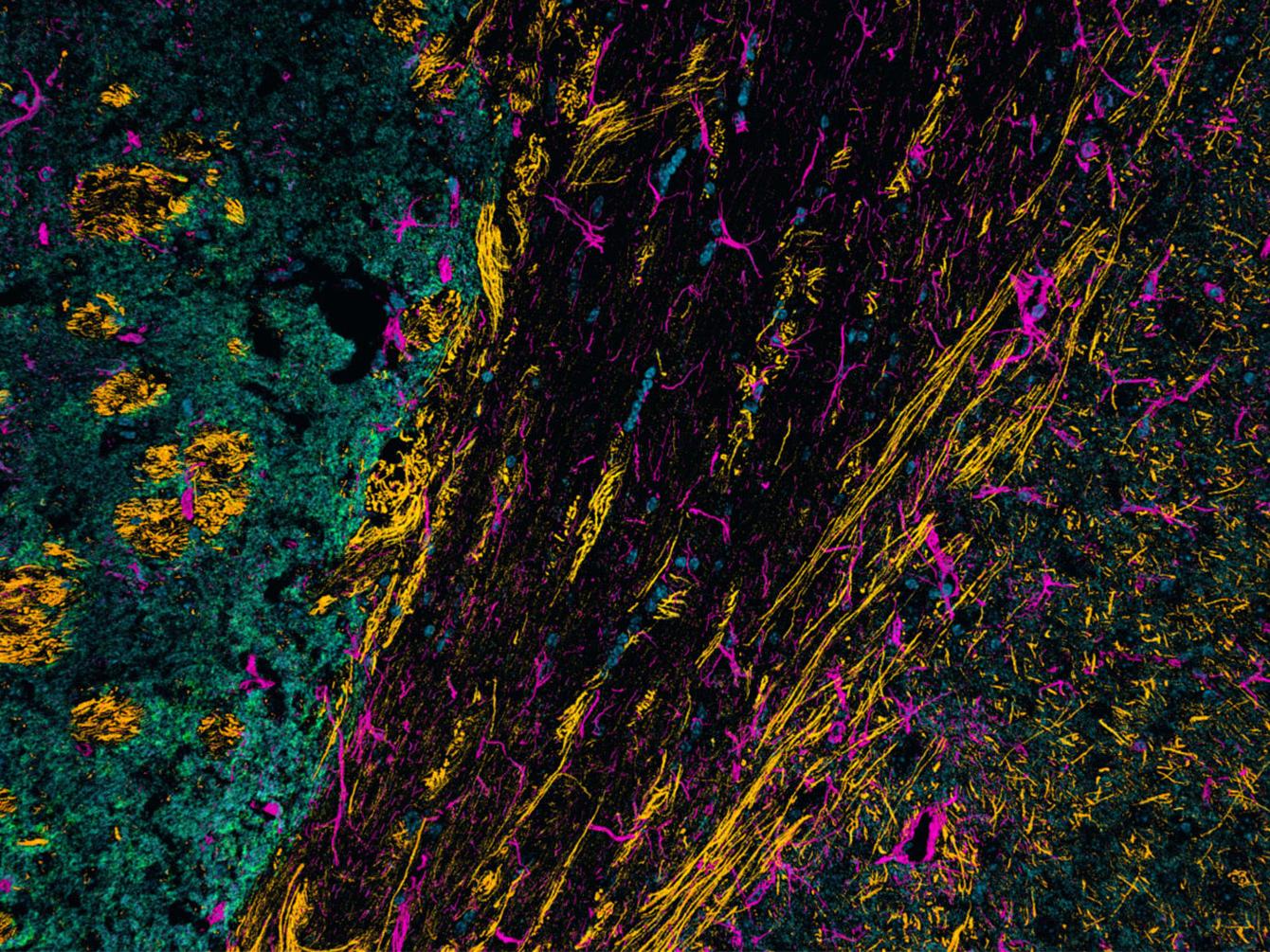
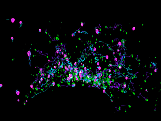
Neuron Tracing
In this series "From Image to Results", explore various case studies explaining how to reach results from your demanding samples and acquired images in an efficient way. For each case study, we highlight different samples, imaging systems, and research questions. In this case study, we trace neurons.


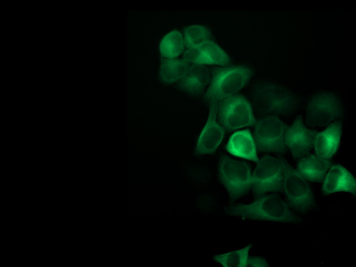

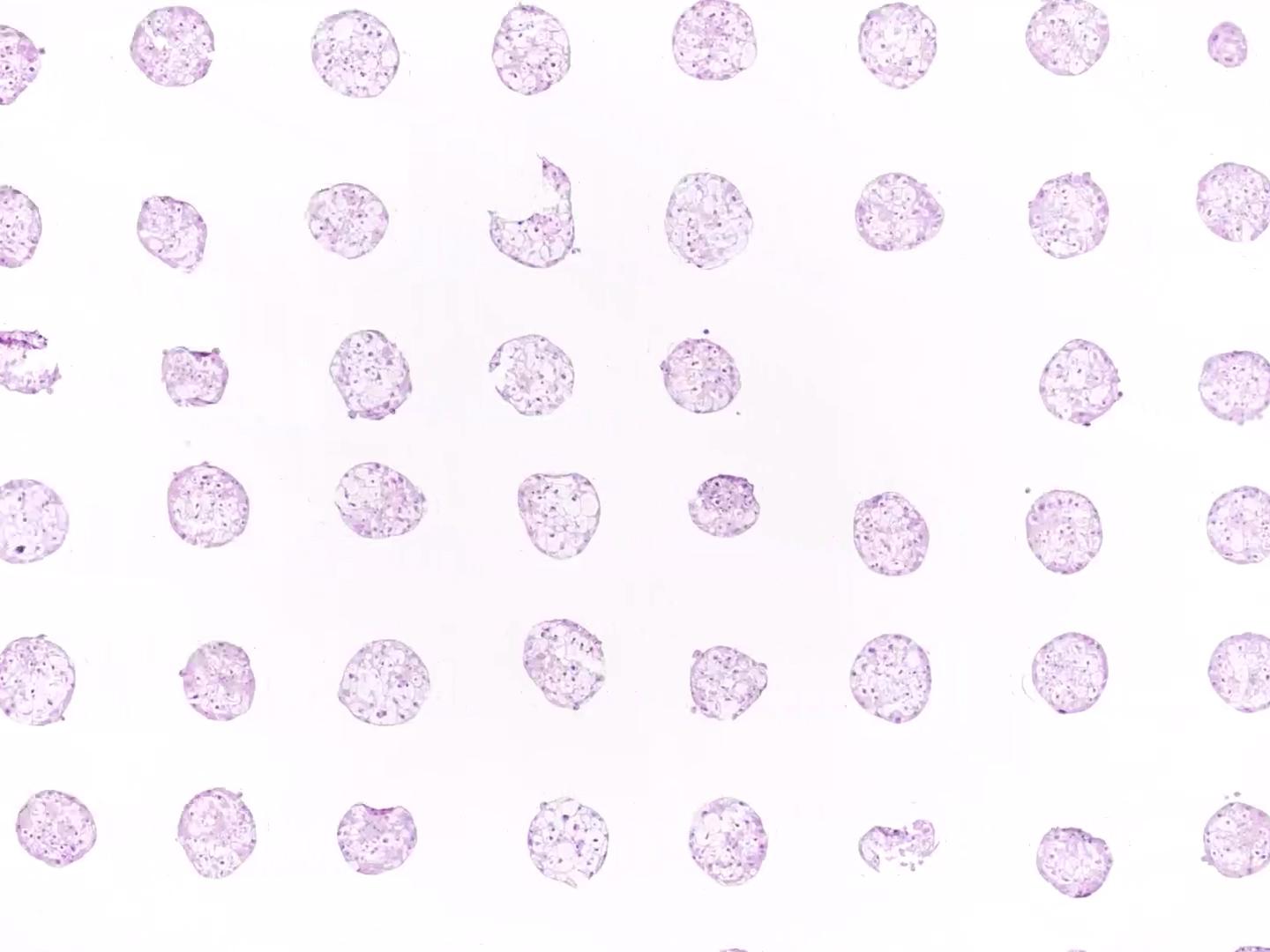
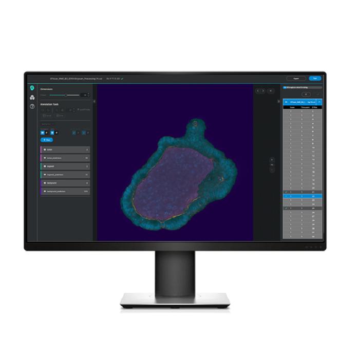
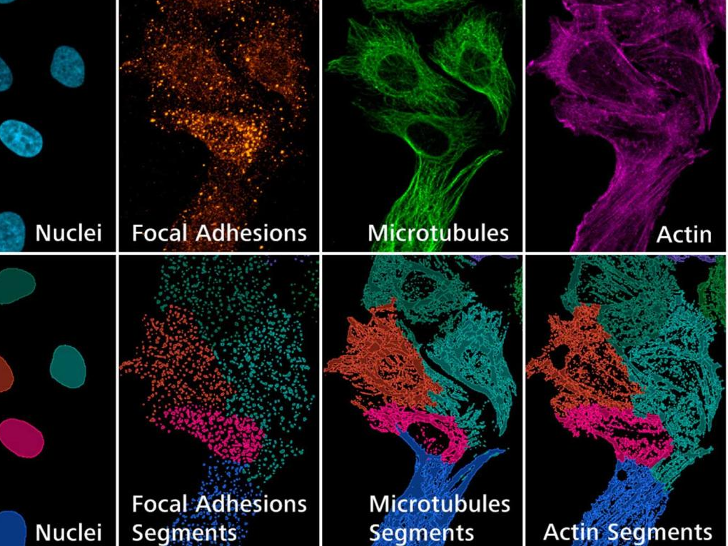
Leveraging High-Content Screening and AI Analysis for Cytotoxicity Assays in Biopharma Research
This approach enhances the efficiency and precision of cytotoxicity studies and facilitates the identification of optimal treatment strategies. This application note demonstrates how this methodology can transform drug development processes and contribute to the advancement of targeted therapies in the biopharmaceutical industry.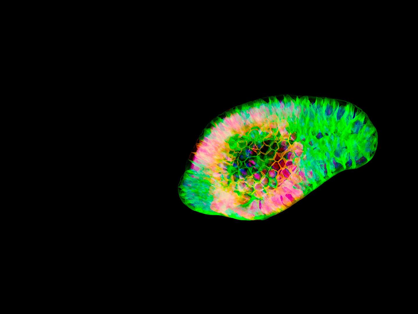

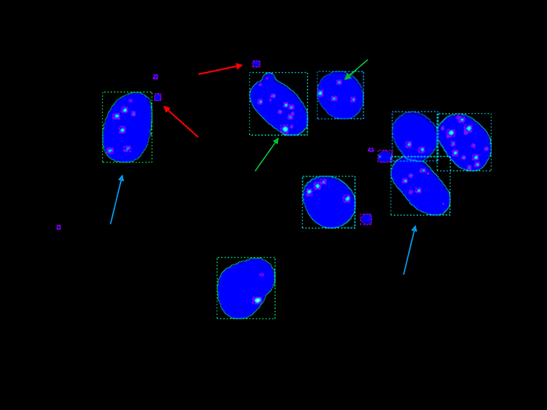

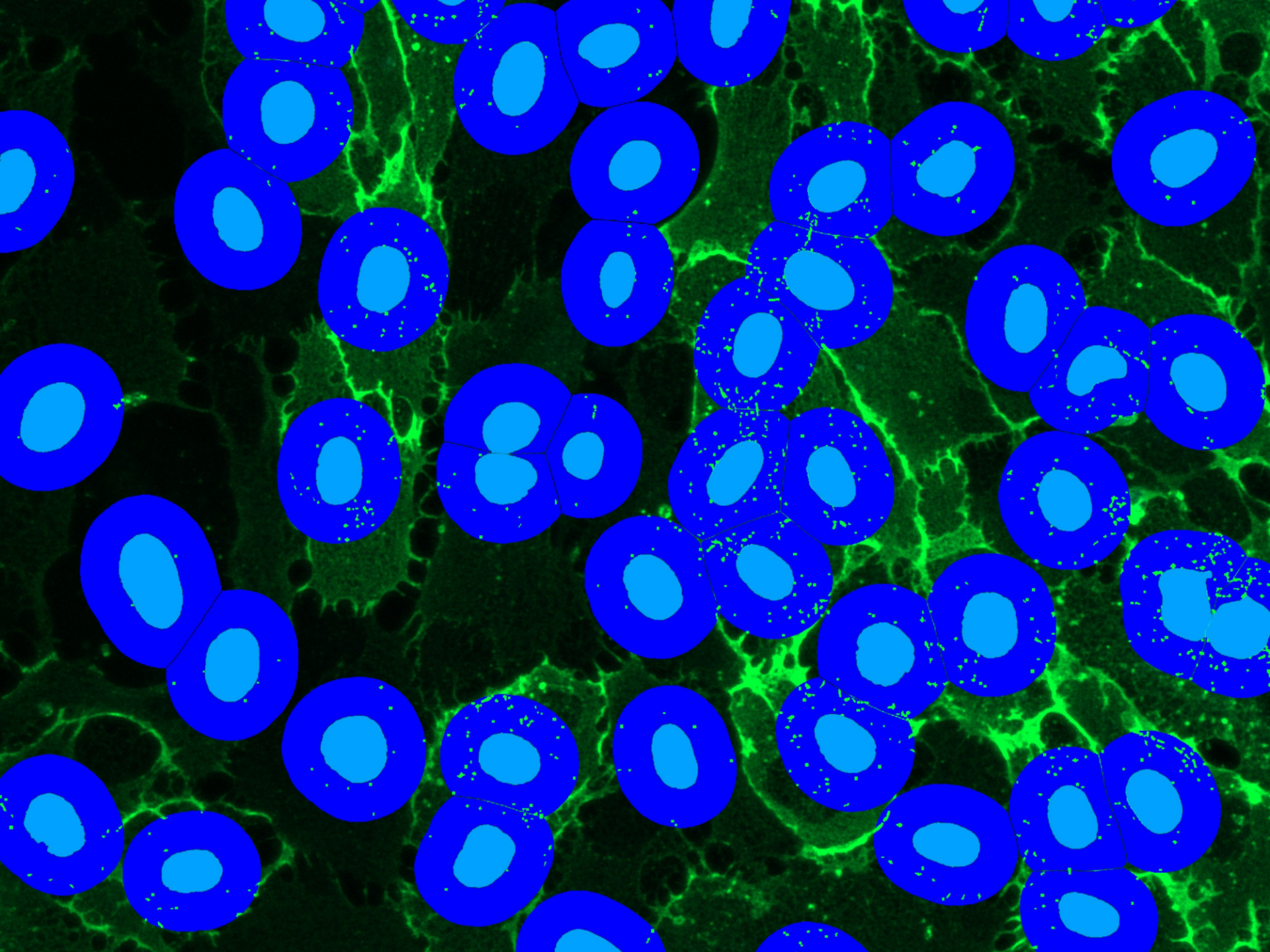
High Content Imaging for Genotoxicity
Key learnings from this episode explores: how to perform the general analysis with one drug and how to automate imaging and analysis procedures, to the point that the whole process can be scaled easily to perform high-content screening (HCS) or be adapted to other complex experimental settings.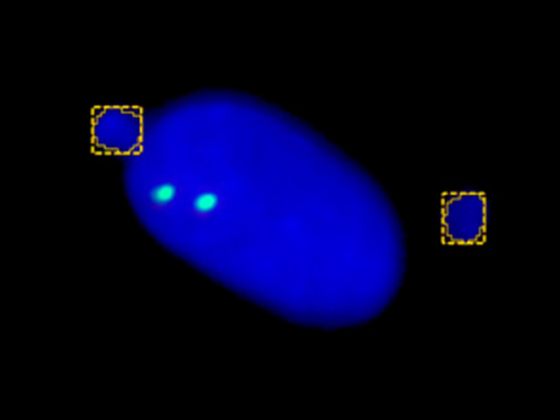


Analyzing Influenza A Virus Entry Through Lattice Light Sheet Microscopy
A research group led by Prof. Allen Liu at the University of Michigan, USA, used ZEISS Lattice Lightsheet 7 to study cellular entry mechanisms of this virus. They focused on the membrane-bending protein epsin. Their findings support the hypothesis that epsin plays a biomechanical role in IAV entry.